- WB
- IHC
- IF/ICC
- Cites

.
Bands result from membrane strip incubation.")
. Green - AF0155")


 and mouse anti-beta tubulin Ab(T0023 1:200) for 1 hour at 37°C. An AlexaFluor594 conjugated goat anti-rabbit IgG(H+L) Ab(Red) and an AlexaFluor488 conjugated goat anti-mouse IgG(H+L) Ab(Green) were used as the secondary antibody.
The nuclear counter stain is DAPI(blue).")
 C6 cells were incubated with FGF2 (10 ng/mL) for different durations. (B, C) Before incubation with FGF2 (10 ng/mL) for 15 min (B) or 24 h (C), cells were pretreated with the FGFR kinase inhibitor SU5402 (SU; 25 lM, 5 min) or the ERK inhibitor U0126 (U0; 10 lM, 1 h). Phosphorylation of FRS2a or ERK1/2 (A and B, respectively), and EAAC1 expression (C) were analyzed by Western blots. Data are shown as mean ± SEM from three independent experiments. * p < 0.05; **p < 0.01 versus the control group. # p < 0.05; ##p < 0.01 versus the FGF2 alone group.")
, U0126 (U,10 μM) for one hour and then incubated with OBHS (1 μM) or RAL (1 μM) for 45 min, after Aβ treatment for an additional 24 h. Cell viability was measured by MTT method. b,c C6 cells were preincubated with PI3K/Akt inhibitor LY (b) or ERK inhibitor U (c) for one hour, followed by OBHS (1 μM) or RAL (1 μM) treatment for 15 min. d GPER1 antagonist G15 (10 μM) was added to the media for 24 h, before OBHS (1 μM) or RAL (1 μM) treatment for 15 min of C6 cells. The proteins were analyzed by the western blot method. The data were shown as the mean±SD from three independent experiments.")
 IGF-1R, CDC42, p85α, p-ERK and ERK mRNA levels were determined by qPCR in ER-positive cells transfected with a miR-29a mimic or miR-29a inhibitor. (B, C) Western blotting analysis was performed to monitor IGF-1R, CDC42, p85α, p-ERK and ERK expression in ER-positive cells transfected with a miR-29a mimic or miR-29a inhibitor. The results showed that ERK, CDC42 and p85α are the target genes of miR-29a; however, miR-29a promotes breast cancer cell growth and proliferation mainly by activating ERK phosphorylation.")

 The effect of miR-377 overexpression on inflammation-related gene expression. *P < 0.05, **P < 0.01 vs. NC 0.1% BSA; ##P < 0.01 vs. NC 10 ng/ml TNFα; ns, not signifcant (n = 3). (B) The effect of miR-377 inhibition on inflammation-related gene expression under conditions of TNFα-induced insulin-resistance. ##P < 0.01 vs. NC 10 ng/ml TNFα(n = 3). (C) The effect of miR-377 overexpression on JNK phosphorylation under conditions of TNFα-induced insulin-resistance. *P < 0.05 vs. NC without insulin; #P < 0.05 vs. NC with insulin (n = 3). (D and E) The effect of miR-377 overexpression on AKT and ERK phosphorylation. *P < 0.05 vs. NC with 0.1% BSA and insulin; #P < 0.05 vs. NC with 10 ng/ml TNFα and insulin (n = 3). (F) The effect of miR-377 inhibition on JNK phosphorylation under conditions of TNFα-induced insulin-resistance. *P < 0.05 vs. NC without insulin; #P < 0.05 vs. NC with insulin (n = 3). (G and H) The effect of miR-377 inhibition on AKT and ERK phosphorylation. *P < 0.05 vs. NC with 0.1% BSA and insulin; #P < 0.05 vs. NC with 10 ng/ml TNFα and insulin (n = 3).")

 Molecular docking results for TP and Pyr. (B) Thymine concentration of NCI-H460 cells treated with 5-UIR, Pyr, and MTX. Intercellular thymine concentration was used as an index of TP activity. (C) Interaction between Pyr and TP verified through Biacore assay (Kd=6.19±0.78 μM). (D) Pyr can inhibit the TP-induced migration of cancer cells. Wound-healing assay was used in this experiment. (E) Pyr can inhibit the TP-induced invasion of cancer cells. Transwell chambers were utilized for the invasion assay, and images were obtained under 200× magnification. (F) E-cadherin, vimentin, MEK2, and ERK2 expression levels of NCI-H460 cells in different treatment groups. The GAPDH blot served as the loading control. Data are presented as the means of three experiments, and error bars represent the standard deviation (*P < 0.05, **P < 0.01)")
 The
effect of SCVP-1 on the level of phosphorylation of ERK1/2, p38 and JNK1/2 in RAW264.7 cells
by Western blotting. (B) Relative quantitative analysis of p-p38/p38, p-ERK/ERK and p-JNK/JNK
protein phosphorylation levels. (C-F) Effect of specific MAPKs inhibitors (ERK1/2 inhibitor:
CI-1040, p38 inhibitor: SB239063 and JNK1/2 inhibitor: SP600125) on SCVP-1/LPS-induced NO,
IL-1β, IL-6 and TNF-α production of RAW264.7 cells. The results were expressed as means ± SD
(n = 3). (B) *p<0.05, **p<0.01 vs. control group. (C-F) *p<0.05, **p<0.01 vs. without
anti-MAPKs antibodies")

, dopamine receptor D2 (DRD2), tyrosine hydroxylase (TH), and dopamine transporter(DAT) in the striatum. After pretreated with MPTP for seven days, the C57BL/6 mice were administrated with MA or different concentrations of OP for the followed seven days.(A): Original bands of ERK1/2, DRD2, TH, DAT, and β-actin.")
 The levels of cyclin A, cyclin B, cyclin D1, and CDK2 were reduced in A375 cells transfected with OVOS2-shRNA; (b) The downregulated expression of N-cadherin accompanied with the upregulated expression of E-cadherin and β-catenin were observed in A375 cells transfected with OVOS2-shRNA; (c) The expression of p-FAK, p-AKT, and p-ERK were reduced in A375 cells transfected with OVOS2-shRNA; (d) The increased production of MMP-2 was observed in A375 transfected with OVOS2-shRNA; (e) GAPDH was used as the reference.")

 After co‑culture with colorectal cancer cells, IL‑6, IL‑8, p‑JNK, and p‑ERK levels in TAMs were examined using western blot assays.In (B) LoVo‑P and (C) HT29‑NC cells, IL‑6 and IL‑8 expression was detected after the addition of an inhibitor to p‑JNK (SP600125; 2.5 and 5 nM) or p‑ERK (SCH772984; 4 and 8 nM) into the co‑culture system with LoVo‑P cells.")
 The protein expression of SMARCE1, ERK1/2, p-ERK1/2, MMP9, and cyclin D1 was examined through western blotting. β-actin was used as control.")
.")
. B. The mRNA expression level of Sp1 in myeloma cell lines among different group (Sp1-siRNA: p = 0.0014; pcDNA3.1-Sp1:p = 0.0001; pcDNA3.1- p300: p = 0.0006). C. The mRNA expression level of p300 in myeloma cell lines among different group (p300-siRNA: p = 0.0012; pcDNA3.1-Sp1: p = 0.0003; pcDNA3.1- p300: p = 0.0002). D. The protein level of Sp1, p300, IQGAP1, ERK1/2 and p-ERK1/2 in myeloma cell lines among different group.")
.")
: Representative immunoblots for PKC, ERK1/2, p-ERK1/2, and tubulin in the HIP region; Levels of PKC and ratio of p-ERK1/2/ERK1/2 in the HIP of rats in each group")
 The phosphorylation of c-Met and downstream effector molecules Akt and Erk was inhibited by CM 7 in MKN-45 cells. MKN-45 cells were treated with the indicated concentrations of peptide CM 7 for 24 h, and the lysate was subjected to Western blot.")
 Representative western blots and quantitative analysis of (B) TGF‑β1/β‑actin, (C) p‑Smad2/3/Smad2/3 and (D) p‑ERK1/2/ERK1/2 levels in rats.")
 and Y5 (S25585, 1 μM concentration) receptor antagonists block the effect of NPY (25 nM) on ERK1/2 phosphorylation in BRL3A hepatocytes. The results are presented as the percentage change in protein intensity compared with the control plus DMSO group and normalized to 100%.")
-induced HepG2 cell death is associated with acid sphingomyelinase (ASM) and extracellular signal-regulated protein kinase(ERK). HepG2 cells were divided into four groups: control (dimethyl sulfoxide), U0126(25 μM), DETA–NO (200 μg/mL) and U0126+DETA–NO groups. After treatment for the indicated times, a apoptosis was analysed via fow cytometry. bThe protein levels of ASM and pERK/ERK were assayed via western blotting. ***P<0.001,compared with the control")
 Western blot analysis of the protein levels of p-Akt, p-Smad3, p-p38 and p-Erk1/2 expression.")
. B‐E, The ERK, P38, JNK, and their phosphorylation forms protein levels of
EA.hy926 cells treated with culture supernatants of HK‐2 cells were measured by western blot (n = 6 for each group). F,G, The eNOS and p‐eNOS
protein levels of EA.hy926 cells treated with culture supernatants of HK‐2 cells were measured by western blot (n = 6 for each group). Data are
presented as mean ± SEM. *P < .05 and **P < .01")
 means
significantly different from the Parkinson disease (PD) control group,
(c) means significantly different from the standard treatment (L-dopa/
carbidopa) control group, (d) means significantly different from inosine
low-dose treatment group, and (e) means significantly different from
inosine medium-dose treatment group at p < 0.05
compared with the standard treatment L-dopa/carbidopa, against
rotenone-induced Parkinson disease (PD) in experimental mice
regarding striatal total and phosphorylated extracellular signal-regulated
kinase 1/2 (tERK and pERK). Each value represents the mean value of 6
measurements ± standard error of the mean (SEM), where (a) means")
 AMPK activation, (B) ERK
activation, (C) JNK activation, and (D) P38 activation. The blots showed representative samples. All samples derived from the same experiment and that blots were
processed in parallel. AMPK and p-AMPK were normalized to the expression of GAPDH on the same membrane. Each phosphoprotein was normalized to the
expression of the corresponding total protein of the same sample in Fig. 5B–D. Results are expressed as mean ± S.E.M. (n = 6–8 for each group). *P < 0.05,
**P < 0.01, ***P < 0.001, ****P < 0.0001 according to one-way ANOVA followed by Bonferroni test.")
 Representative images of ERK1/2 phosphorylation in adolescent and adult offspring
of rat dams intravenously injected with poly I:C (10 mg/kg) or saline on E9.5. (B) Statistical analysis of the ratio of ERK1 phosphorylation to total ERK1. The mean
value of the control group was set at 100, and the data for pups from MIA-injected dams were recorded as a percentage of the mean value of pups from saline-treated
mothers. (C) Statistical analysis of the ratio of ERK2 phosphorylation to total ERK2. The mean value of the control group was set at 100, and the data for pups from
MIA-injected dams were recorded as a percentage of the mean value of pups from saline-treated mothers; n = 6 for each group. Data are means ± SEM. *P<0.05,
**P<0.01 (two-tailed Student's t-test).")
, p-ERK (B), p-JNK (C), p-p38 (G), p-STAT3 (D), p-p65 (E), SIRT1 (F), and PGC-1α (H) were
determined by western blotting in lung tissues. The lung tissues were dissected 24 h after LPS challenge and the levels of protein expressions were evaluated by
densitometric analysis of the blots. The blots showed representative samples. All samples derived from the same experiment and that blots were processed in parallel.
SIRT1 and PGC-1α were normalized to the expression of GAPDH on the same membrane. Each phosphoprotein was normalized to the expression of the corresponding
total protein of the same sample. Results were expressed as mean ± SD (n = 6 for each group). *P < 0.05, **P < 0.01, ***P < 0.001, ****P < 0.0001 according
to one-way ANOVA followed by Tukey’s multiple comparisons test.")
 western blot analysis. (B) Quantification of western blotting results. ***P<0.001 vs. the control group. HYSA, hydroxysafflor yellow A; p, phosphorylated.")
 Western blot analysis of p-ERK1/2, total ERK1/
2, p-P38 MAPK, total P38 MAPK, p-JNK, and total JNK. These results were used
to calculate (B) p-ERK1/2/β-actin and total ERK1/2/β-actin ratios, (C) p-P38
MAPK/β-actin and total P38 MAPK/β-actin ratios, (D) p-JNK/β-actin and total
JNK/β-actin ratios. The differences were statistically significant (p < 0.05).
Asterisk indicates significant difference in comparison to untreated cell")
 expression through the ERK signaling pathway in H2228 cells. A, Phosphorylation level of ERK1/2 protein after treatment using ERK inhibitor U0126. GAPDH was utilized to verify equal loading. B, Relative expression level of PD‐L1 mRNA after treatment using ERK inhibitor U0126. C, Phosphorylation level of ERK1/2 protein after transfection of ERK overexpression plasmid (OE). GAPDH was utilized to verify equal loading. D, Protein expression level of PD‐L1, which was stably transfected with control vector plasmid and ERK overexpression plasmid for 24 h. *P < .05, **P < .01. p‐, phosphorylated")
 Western blot analysis of AKT/ERK1/2, G1-phase checkpoint and EMT molecules. (A) The representative western blot images. (B) The relative level of p-AKT (T308)/AKT, p-AKT (S473)/AKT and p-ERK1/2/ERK1/2 as well as p21, p27, cyclin D1, cyclin E1, CDK2, CDK4, E-cadherin, N-cadherin, vimentin, Snail and Slug in A549-SIRT7 (A549-mock served as a control) or H292-shSIRT7 (H292-shcontrol served as a control) NSCLC cells. *P<0.05, Student's t-test, n=6 per group. (C) IHC analysis of AKT/ERK1/2, G1-phase checkpoint and EMT molecules in xenograft tumor tissues. The representative IHC images are presented. SIRT7, sirtuin 7; EMT, epithelial-mesenchymal transition; NSCLC, non-small cell lung cancer; AKT, protein kinase; ERK1/2, extracellular signal-regulated kinase 1/2; CDK, cyclin-dependent kinase; IHC, immunohistochemistry.")
 Western blot analysis of AKT/ERK1/2, G1-phase checkpoint and EMT molecules. (A) The representative western blot images. (B) The relative level of p-AKT (T308)/AKT, p-AKT (S473)/AKT and p-ERK1/2/ERK1/2 as well as p21, p27, cyclin D1, cyclin E1, CDK2, CDK4, E-cadherin, N-cadherin, vimentin, Snail and Slug in A549-SIRT7 (A549-mock served as a control) or H292-shSIRT7 (H292-shcontrol served as a control) NSCLC cells. *P<0.05, Student's t-test, n=6 per group. (C) IHC analysis of AKT/ERK1/2, G1-phase checkpoint and EMT molecules in xenograft tumor tissues. The representative IHC images are presented. SIRT7, sirtuin 7; EMT, epithelial-mesenchymal transition; NSCLC, non-small cell lung cancer; AKT, protein kinase; ERK1/2, extracellular signal-regulated kinase 1/2; CDK, cyclin-dependent kinase; IHC, immunohistochemistry.")
. *P < .05, **P < .01 compared with the CON group; #
P < .05, ##P < .01 compared with the THP group. CON, control;
mRNA, messenger RNA; qPCR, quantitative real‐time polymerase chain reaction; RAP1/ERK, RAP1‐member of RAS oncogene family/extracellular
regulated protein kinases; RUT, rutin; THP, pirarubicin")
 NB4 cells were stimulated with 0, 10, 20, and 30 μM HNK for 24 h, and the expression of MAPK signaling pathway related proteins was determined. The cells were divided into four groups: Solvent, HNK(30 μM), HNK + U0126/SP600125/SB203580, U0126/SP600125/SB203580.")
 and statistically analyzed with Image J software (B, C).")
 and
mDCs (c, d) isolated from mice fed with diets containing low
(0.08 ppm), medium (0.25 ppm) or high (1 ppm) selenium for 8 weeks Data are presented as mean ± SD (n = 3). *P < 0.05; **P < 0.01; ***P <
0.001; low, medium and high respectively represent DCs isolated from
mice fed with different concentrations of selenium")
 NR activated the JAK2/Stat3 pathway by increasing the levels of p-JAK2 and pStat3. Relative levels of p-JAK2 and p-Stat3 were normalized to the JAK2 and Stat3 levels, respectively. (c-d) NR inhibited the H2O2-induced phosphorylation of MAPK.
Relative levels of p-JNK and p-ERK 1/2 were normalized to the JNK and ERK 1/2 levels, respectively (**p < .01, ***p < .001, and ****p < .0001 compared to the H2O2
group).")
 Expression of colonic phosphorylated ERK1/2 and phosphorylated p38 MAPK in colon tissue as
determined by western blot. (B) Expression of NF-κB in colon tissue as determined by western blot.")

, Ts-ML-ES (25 μg/ml), heat-inactivated Ts-ML-ES (25 μg/ml), or PMSF-Ts-ML-ES (25 μg/ml). β-actin was used as the internal control. Representative blots of three independent experiments are shown.")
 Representative images of Western blot. (B–I) quantification of the Western blot data by densitometric analysis and normalization to GAPDH (n = 3 independent experiments). **P < 0.01 vs model group.")

 and C/EPBα (b) were examined on days 1–3, respectively. Cells in 10% FBS were used as control group. N = 3. *, p < 0.05, vs. 10% FBS group; **, p < 0.01, vs. 10% FBS group; ##, p < 0.01, vs. 2% FBS group; &&, p < 0.01, vs. 10% FBS group on day 1. B. ADSCs were treated with 2% FBS and/or d-ECM. Cells without any treatment were set as control. For d-ECM treatment group, a copy pretreated with 50 μM PD98059 was established to inhibit ERK1/2 signalling (a). Afterwards, the phosphorylation of ERK1/2 (b), and the expression levels of PPARγ (c) and C/EPBα (d) were examined by Western blotting analysis. Equal loading of proteins was demonstrated by GAPDH. N = 3. *, p < 0.05, vs. 10% FBS group; **, p < 0.01, vs. 10% FBS group; ##, p < 0.01, vs. 2% FBS group; $$, p < 0.01, vs. 2% FBS + d-ECM group. P-ERK1/2, phospho-extracellular signal-regulated kinase1/2; ERK1/2, extracellular signal-regulated kinase1/2; PPARγ, peroxisome proliferators-activated receptor γ; C/EPBα, CCAAT enhancer-binding proteins")
.")
 and statistically analyzed with Image J software (B, C). Data are presented as mean±SD of 3 independent experiments. * P<0.05, ** P<0.01, *** P<0.001, compared with the control group.")

 Expression of TLR4, phosphorylated p38 (P-p38), p38, P-ERK1/2, ERK1/2, P-JNK, JNK, P-p65, p65 was shown. (B) The analysis of grey scale value. Con was control group, M was model group, H was the high dose OPA group, L was low dose OPA group, P was positive group. Data were expressed as the mean ± SD, the statistical analyses were done with t-test; *** p < 0.001, ** p < 0.01, * p < 0.05 compared with the model group; ### p < 0.001, ## p < 0.01, # p < 0.05 compared with the control group.")
 Representative safranin O/fast green‐stained cartilage histological images and OARSI scores for all groups. (B) Western blotting analysis of MMP‐3, MMP‐13, ADAMTS‐5, ICAM‐1 and β‐actin proteins in synovial tissues for all groups. (C) Western blotting analysis of p‐ERK, ERK and β‐actin proteins in synovial tissues for all groups. *P < 0.05, ****P < 0.0001. n = 6. Ctrl, control; DM‐OA, diabetes mellitus‐osteoarthritis; LV‐NC, negative control lentivirus transfected; LV‐PSTPIP2, PSTPIP2 overexpression lentivirus transfected.")

 knockdown inhibited the activation of the mitogen-activated protein kinase (MAPK) pathway 3 days after myocardial infarction (MI). The expression levels of p-JNK, c-Jun N-terminal kinase (JNK), p-p38, p-38, p-ERK, and ERK were determined using western blot analysis. β-actin served as the loading control.")

The markers of MEK/ERK signaling pathway including p-ERK1/2, ERK1/2, p-MEK, MEK, and Myc were detected in cells transfected with
AQP5-overexpressed plasmid. (B, C) After AGS (B) and GES-1 (C) cells infection with H. pylori and treatment with AQP5 siRNA, the markers
of MEK/ERK signaling pathway was examined by Western blot. (D) Downstream genes of MEK/ERK signaling pathway including ELK1, Myc,
and c-FOS were detected by RT-PCR analysis in AGS cells treated with H. pylori and AQP5 siRNA. (E) Western blot analysis was performed
to detect the expression of EMT markers in cells following H. pylori infection and treatment with ERK inhibitor U0126. (F) Schematic for
the proposed mechanism of action of AQP5 in H. pylori CagA-associated gastric tumorigenesis. CagA+
H. pylori delivers CagA into gastric
epithelial cells and then induces oncogene AQP5 expression. Subsequently, the MEK/ERK signaling pathway is activated, which induces the
EMT program and thereby further promoting gastric tumorigenesis")
 Compound PIF_9 inhibited the activation of MAPK signaling pathway. RAW264.7 cells in a 6-well plate cultured in serum-free medium were pretreated with compound PIF_9 for 1 h, then co-treated with LPS (100 ng/mL) for 2 h. (C) RAW264.7 cells were pretreated with compound PIF_9 for 1 h, then co-treated with LPS (100 ng/mL) for 24 h. Cellular ROS levels was determined by detection of DCF fluorescence intensity as described in Materials and Methods. (D,E) RAW264.7 cells were pretreated with compound PIF_9 for 1 h, then co-treated with LPS (100 ng/mL) for 24 h. After treatment, cellular protein was extracted and protein expression levels of p-JNK, JNK, p-p38, p38, p-ERK, ERK (A,B), and SOD1 (D, E) were analyzed by Western blot, followed by quantitative analysis of band density as described in Materials and Methods. ##, p < 0.01, ###, p < 0.001, vs. Control; *, p < 0.05, **, p < 0.01, ***, p < 0.001 vs. LPS-treated group; ns, no significant difference; ND, no detection (n = 3).")
 Luciferase assays of CAGA-NIH3T3 cells. Cells were pretreated with Remdesivir (0–50 μM) for 30 min and then incubated with TGF-β1 (5 ng ml−1) for 24 h, then analyzed with luciferase assay. SB431542 is a TGF-β1/Smad pathway inhibitor and serves as a positive control (B) NIH-3T3 cells were co-treated with TGF-β1 (5 ng ml−1) and Remdesivir (12.5, 25, 50 μM) for 1 h. P-Smad3 and Smad3 were assessed using western blot. GAPDH was used as the internal control (C) PPF cells were co-treated with TGF-β1 (5 ng ml−1) and Remdesivir (12.5, 25, 50 μM) for 1 h. P-Smad3 and Smad3 were assessed using western blot. GAPDH was used as the internal control (D) NIH-3T3 cells were co-treated with TGF-β1 (5 ng ml−1) and Remdesivir (12.5, 25, 50 μM) for 1 h and the phosphorylation levels of P-38, JNK, ERK and Akt were analyzed by Western blot. β-tubulin was used as a loading control in grayscale analysis. Scale bar = 60 μm. Data was presented as the means ± SD, n = 3. * p < 0.05, ** p < 0.01, *** p < 0.001, **** p < 0.0001.")
. The phosphorylation levels of (B) p70S6K and 4E-BP1; (C) AMPK, ACC, and ULK1; (D) ERK1/2; (E) p38MAPK; and (F) eEF2 are shown. The phosphorylation is normalized to the total protein expression. The β-tubulin content in the lysate was measured as a loading control (G). The time course of these experiments is shown in the upper region. DM: differentiation medium, DMEM: Dulbecco’s modified Eagle’s medium, and w/o AA: without amino acids. Data are displayed as the means ± SD, and n = 4 for each group in all bar graphs. * p < 0.05 and ** p < 0.01 vs. the vehicle-treated group.")
 AQP3 expression; (C,D) VIP, VIPR2, cAMP and PKA expression; (E,F) MEK1/2, p-MEK1/2, ERK, p-ERK, p-p38, p38, MSK1, p-MSK1, CREB, p-CREB, and P300/CBP expression. Data are shown as mean ± SD (n = 3). # p < 0.05, ## p < 0.01 versus control group; * p < 0.05, ** p < 0.01 versus 5-FU group.")
 Experimental procedure of DARTS with TSG. (B) Coomassie SimplyBlue staining of the cerebral lysates subjected to proteinase K digestion (n = 3). (C) SDS-PAGE analysis of the cellular lysates subjected to proteinase K digestion with A1R antibody staining (n = 3). (D) The phosphorylation of ERK1/2 in HEK293T cells without A1R was detected by Western blot after 24 h incubation with adenosine (ADO, 8 µM) and TSG (4 µM and 8 µM). (E,F) The phosphorylation of ERK1/2 in HEK293T/A1R cells and SH-SY5Y cells was detected by Western blot after 24 h treatment of ADO (8 µM), DPCPX (5 µM) and TSG (4 µM and 8 µM). The gray value was analyzed by ImageJ software. All the values were expressed as mean ± SD, n ≥ 3. (G) The heart rate and blood pressure of rats 14 days before TSG treatment and 21 days with TSG treatment, respectively. TSG was treated for rats for 21 days with 40, 20 and 10 mg/kg (s.c., twice a day) corresponding to the TH, TM and TL groups. Each point represents the mean value of blood pressure and heart rate of rats each day in the same group. The observed vertical bar represents the mean ± SD of the blood pressure and heart rate at 14 days and 21 days, respectively. * p < 0.05, and *** p < 0.001, other groups vs. Control group.")

The levels of pro-inflammatory cytokines TNF-α and IL-6 in BALF were
measured by ELISA assay. (B) The
expression of Bcl-2 and Bcl-xl were
detected by western blotting. (C) The
expression of MEK, p-MEK (Ser217/
Ser221), ERK, p-ERK (Thr202/Tyr204),
IKK-α/β, p-IKK-α/β (Ser180/Ser181),
IκB-α, p-IκB-α (Ser32/36), NF-κB p65
(cytoplasm), NF-κB p65 (nucleus) were
determined by western blotting. Data
were shown as means ± SD. n = 6. *p <
0.05 vs. sham. #p < 0.05 vs. CLP. ERK,
extra-cellular signal-regulated kinase;
NF-κB, nuclear factor-kappa B; TNF-α,
tumor necrosis factor-α; IL-6, interleukin-6; BALF, bronchoalveolar lavage
fluid; CLP, cecal ligation and puncture.")
 Western blot analysis of NF-κB, p-iκB, p-NF-κB, p-ERK1/2, and p-P38 in cells treated with RANKL (50 ng/mL) for different amounts of time (0, 10, 20, 30, and 60 min) in the presence or absence of peiminine (10 μmol/L). Quantitative analysis of the differential expression levels of (B) NF-κB (n= 3), (C) p-NF-κB (n= 4), (D) p-iκB (n= 4), (E) p-ERK1/2 (n= 3), (F) p-P38 (n= 3). β-tublin was selected as the control protein for the normalization of p-iκB, NF-κB, p-NF-κB, and the ERK1/2 for p-ERK1/2, the P38 for p-P38. *P < 0.05, **P < 0.01, ***P < 0.001.")
.")
: representative immunoreactive bands of ODC, VEGF, MMP-9, GAPDH, total and phosphorylated Erk1/2 using specific antibody. (B–E): representative quantification of ODC, phosphorylated Erk1/2, VEGF, MMP-9. (F): determination of MMP-9 activity. * p < 0.05, ** p < 0.01, *** p < 0.001 compared with MCF-7/ADR cells, ## p < 0.01, ### p < 0.001, comparison between MCF-7/ADR group and MCF-7 group.")
 AGS cells were transfected with control-siRNA or HRC-siRNA and stained with Fluo‑4/AM. (B) Knockdown of HRC reduced the intracellular Ca2+ concentration in AGS cells. (C) MKN-45 cells were transfected with control-siRNA or HRC-siRNA and stained with Fluo‑4/AM. (D) Knockdown of HRC reduced the intracellular Ca2+ concentration in MKN-45 cells. (E-H) Levels of p-c-Raf, c-Raf, p-MEK, MEK, p-ERK, ERK, E-cadherin, N-cadherin, Vimentin, Snail and Slug proteins in AGS and MKN-45 cells were analyzed by western blot. HRC, histidine-rich calcium binding protein; siRNA, small interfering RNA; c-Raf, Raf-1 proto-oncogene, serine/threonine kinase; p-c-Raf, phosphorylated c-Raf; MEK, MAPK/ERK kinase 1; p-MEK, phosphorylated MEK; ERK, extracellular regulated kinase; p-ERK, phosphorylated ERK; CaM, calmodulin. Data are shown as the mean ± SD; ns: no significant difference; *P < 0.05, **P < 0.01, ***P < 0.001, based on Student's t test.")
 MDA-MB-231 cells and adipocytes were co-cultured in the Transwell system, and selective pathway inhibitor (A) or ERK1/2 downstream transcription factor inhibitor were added to detect expression (B) and secretion (C) of LIF. (D) The protein expression level in adipocytes was analyzed by western blot. (E-G) LY3214996 (E), BAY 11-7082 (F), or Stattic (G) were added to the co-culture system of MDA-MB-231 cells and adipocytes. The protein expression level in adipocytes was analyzed by western blot. (H) The adipocytes were labeled with specific antibodies of p-ERK1/2, NF-κB p65 and Stat3, and the expression and localization of the three in adipocytes were observed under a fluorescent confocal microscope, scale bar: 20 μm. Typical microscopic fields and blots are shown and quantitative data are presented as the mean ± SD from at least three independent experiments. #p < 0.05, ##p < 0.01, ###p < 0.001, Co-culture + DMSO VS. Control + DMSO; *p < 0.05, **p < 0.01, ***p < 0.001, Co-culture +inhibitors VS. Co-culture + DMSO.")
 MDA-MB-231 cells and adipocytes were co-cultured in the Transwell system, and selective pathway inhibitor (A) or ERK1/2 downstream transcription factor inhibitor were added to detect expression (B) and secretion (C) of LIF. (D) The protein expression level in adipocytes was analyzed by western blot. (E-G) LY3214996 (E), BAY 11-7082 (F), or Stattic (G) were added to the co-culture system of MDA-MB-231 cells and adipocytes. The protein expression level in adipocytes was analyzed by western blot. (H) The adipocytes were labeled with specific antibodies of p-ERK1/2, NF-κB p65 and Stat3, and the expression and localization of the three in adipocytes were observed under a fluorescent confocal microscope, scale bar: 20 μm. Typical microscopic fields and blots are shown and quantitative data are presented as the mean ± SD from at least three independent experiments. #p < 0.05, ##p < 0.01, ###p < 0.001, Co-culture + DMSO VS. Control + DMSO; *p < 0.05, **p < 0.01, ***p < 0.001, Co-culture +inhibitors VS. Co-culture + DMSO.")
 The transcript levels of PTPRH and EGFR with or without IL-13 stimulation were detected by qRT-PCR. (B) The protein expression in HBECs with or without IL-13 stimulation were detected by Western blotting. (C) The protein expression in HBECs transfected with empty or PTPRH over expression vector were detected by Western blotting. (D) The protein expression in HBECs transfected with empty or PTPRH siRNA were detected by Western blotting. The transcript levels are expressed as log2 transformed and relative to the mean of control group. Data are mean ± SD. *p < 0.05; **p < 0.01.")
 The protein expression of p-p65, p65, p-ERK1/2, ERK1/2, p-p38, p38, p-JNK, and JNK were determined by Western blot and quantified. GAPDH was conducted as a loading control. (c) The nuclei translocation of p65 (red) was detected by the immunofluorescence assay. Nuclei (blue) were stained with DAPI. One-way ANOVA, ##p < 0.01, compared with control cells; $p < 0.05, $$p < 0.01, compared with IL-1β-treated cells.")
 hirudin and 10 μM losartan for 24 h, respectively. (A) The expression of STAT3, MAPK1 and IL-6 were analyzed by qRT-PCR, n = 6. (B) The IL-6 in cell supernatant was assayed by ELISA, n = 5. (C) Western blot for the expression of proteins including STAT3, p-STAT3, MAPK1, p-MAPK1 and IL-6. (D) Quantified by western blot analysis and normalized to control, n = 3. Bars represent the mean ± SD, *p < 0.05, **p < 0.01.")
 Western blotting was performed to detect the expression of p65 and p-p65 in TNF-α-stimulated FLSs. (B) Expression levels of ERK1/2, p-ERK1/2, JNK, p-JNK, p38 and p-p38. NF-κB, nuclear factor-kappa B; MAPK, mitogen-activated protein kinase. The data represent the mean ± SD (n=3). ##P<0.01 vs. Control; **P<0.01 vs. TNF-α+NC siRNA.")

 Western blot analyses and quantitative data. The impact of FPR2 activation by WKYMVm on phosphorylation of p38, ERK1/2, NF-κB p65, and IκBα protein level in LPS-treated HAPI microglia with or without pretreatment of WKYMVm. (d–f) Western blot analyses and quantitative data for p-p38, p-ERK1/2, p-NF-κB p65, and IκBα from each group in primary microglia. SB203580: p38-specific inhibitor; U0126: ERK-specific inhibitor; BAY11-7082: NF-κB p65-specific inhibitor. ∗p < 0.05 vs. the control group; #p < 0.05 vs. the LPS group.")
 AOPPs (200 µg/mL, 24 h) significantly increased phosphorylation of p38, ERK1/2 and JNK in AF cells. (D–F) p38 MAPK inhibitor (SB202190, 10 µM), ERK1/2 inhibitor (FR180204, 10 µM) and JNK inhibitor (SP600125, 10 µM) AOPPs-induced (200 µg/mL, 24 h) senescence in AF cells. (G–I) Activation of AOPPs-induced (200 µg/mL, 24 h) MAPK phosphorylation was ameliorated by GKT (20 µM) and APO (100 µM). *P < 0.05 compared to the control. ** P < 0.01 compared to the control. #P < 0.05 compared to the AOPPs group. Error bars represent standard errors.")
 qPCR analysis of RAGE, TLR2, and TLR4 mRNA levels in GSCs under hypoxia for 24 h. Data are normalized to normoxia group and represented as mean ± SD Significance was determined by two-tailed unpaired Student’s t test. n = 3 replicates for each group. ∗∗∗, p < 0.001; ∗∗∗∗, p < 0.0001.
(B and C) Western blot and quantitative analysis of TLR2 and TLR4 expression in GSCs with HMGB1 knockdown under hypoxia for 24 h. (B) Representative blots are shown. (C) Data are normalized to shNT group and represented as mean ± SD Significance was determined by two-tailed unpaired Student’s t test. n = 3 replicates for each group. ns, no significance.
(D) The correlation of HMGB1 mRNA expression with AGER (RAGE mRNA) in patients with GBM (IDH wt) from CGGA database using Pearson’s correlation analysis. n = 288, r = 0.4494; ∗∗∗∗, p < 0.0001.
(E and F) Immunofluorescent double staining of HMGB1 (red) and RAGE (green) in human GBM specimens. DAPI stained nuclei (blue). (E) Representative images are shown. Scale bar, 25μm. (F) Quantitative analysis of cells co-expressing HMGB1 and RAGE in total cells. Data are represented as mean ± SD 9 random microscope fields of each GBM specimen (n = 3) were selected.
(G and H) GSEA and heatmap show upregulation of cell cycle progression genes in patients with GBM with higher expression of HMGB1 based on the CGGA dataset. NES, normalized enrichment score. FDR, false discovery rate.
(I and J) Western blot and quantitative analysis of the expression of RAGE-dependent ERK1/2 signaling molecules in GSCs with HMGB1 knockdown under hypoxia for 24 h. (I) Representative blots are shown. (J) Data are normalized to shNT group and represented as mean ± SD Significance was determined by two-tailed unpaired Student’s t test. n = 3 replicates for each group. ∗, p < 0.05; ∗∗, p < 0.01; ∗∗∗, p < 0.001.")
 WB analysis of the AKT/ERK1/2-GSK3β-β-catenin/Snail axis and its regulatory targets in TIPE3 knock-down and TIPE3-overexpressing NSCLC cells. Representative WB images are shown. (B) IF analysis of the AKT/ERK1/2-GSK3β-β-catenin/Snail axis in TIPE3 knock-down and TIPE3-overexpressing NSCLC xenograft tumor tissues. Representative IF images of merge (red, Alexa Fluor 647; green, GFP; and blue, DAPI) (×200) are shown. (C) WB analysis of β-catenin, Snail1, and Slug in TIPE3-overexpressing NSCLC cells after treatment with PI3K or MEK inhibitor and TIPE3 knock-down NSCLC cells after treatment with GSK3β or proteasome inhibitor. Representative WB images are shown. (D) WB analysis of β-catenin, Snail1, and Slug in TIPE3 knock-down NSCLC cells after β-Trcp or STUB1 knockdown. Representative WB images are shown. (E,F) Clinical association of TIPE3 with the AKT/ERK1/2-GSK3β-β-catenin/Snail axis in lung cancer. Representative IHC images of Case 27 (+++, high TIPE3 expression) and Case 34 (-, low TIPE3 expression) lung cancer T tissues (×400) (E) are shown. Percentage of lung cancer T tissue specimens showing high or low TIPE3 expression in relation to the expression level of proteins related to the AKT/ERK1/2-GSK3β-β-catenin/Snail axis (F) is shown. *, P<0.05, Pearson’s χ2 test. (G) A schematic model for the mechanism of action of TIPE3 during NSCLC progression. Upregulation of TIPE3 promotes growth and metastasis in NSCLC cells by activating β-catenin, Snail1, and Slug transcriptional signaling via the AKT/ERK1/2-GSK3β axis. NSCLC, non-small cell lung cancer; WB: western blotting; AKT, protein kinase B; ERK1/2, extracellular signal-regulated kinase 1/2; GSK3β, glycogen synthase kinase 3β; IF, immunofluorescence; GFP, green fluorescent protein; DAPI: 4’,6-diamidino-2-phenylindole; PI3K, phosphatidylinositol 3-kinase; MEK: mitogen-activated protein kinase kinase; IHC, immunohistochemistry; T, lung cancer tumor.")
 WB analysis of the AKT/ERK1/2-GSK3β-β-catenin/Snail axis and its regulatory targets in TIPE3 knock-down and TIPE3-overexpressing NSCLC cells. Representative WB images are shown. (B) IF analysis of the AKT/ERK1/2-GSK3β-β-catenin/Snail axis in TIPE3 knock-down and TIPE3-overexpressing NSCLC xenograft tumor tissues. Representative IF images of merge (red, Alexa Fluor 647; green, GFP; and blue, DAPI) (×200) are shown. (C) WB analysis of β-catenin, Snail1, and Slug in TIPE3-overexpressing NSCLC cells after treatment with PI3K or MEK inhibitor and TIPE3 knock-down NSCLC cells after treatment with GSK3β or proteasome inhibitor. Representative WB images are shown. (D) WB analysis of β-catenin, Snail1, and Slug in TIPE3 knock-down NSCLC cells after β-Trcp or STUB1 knockdown. Representative WB images are shown. (E,F) Clinical association of TIPE3 with the AKT/ERK1/2-GSK3β-β-catenin/Snail axis in lung cancer. Representative IHC images of Case 27 (+++, high TIPE3 expression) and Case 34 (-, low TIPE3 expression) lung cancer T tissues (×400) (E) are shown. Percentage of lung cancer T tissue specimens showing high or low TIPE3 expression in relation to the expression level of proteins related to the AKT/ERK1/2-GSK3β-β-catenin/Snail axis (F) is shown. *, P<0.05, Pearson’s χ2 test. (G) A schematic model for the mechanism of action of TIPE3 during NSCLC progression. Upregulation of TIPE3 promotes growth and metastasis in NSCLC cells by activating β-catenin, Snail1, and Slug transcriptional signaling via the AKT/ERK1/2-GSK3β axis. NSCLC, non-small cell lung cancer; WB: western blotting; AKT, protein kinase B; ERK1/2, extracellular signal-regulated kinase 1/2; GSK3β, glycogen synthase kinase 3β; IF, immunofluorescence; GFP, green fluorescent protein; DAPI: 4’,6-diamidino-2-phenylindole; PI3K, phosphatidylinositol 3-kinase; MEK: mitogen-activated protein kinase kinase; IHC, immunohistochemistry; T, lung cancer tumor.")
 Western blots showing the levels of phosphorylated p38, ERK1/2, and Akt in the sham, 24 h tMCAO, CAY-10444 + 24 h tMCAO, and V+24 h tMCAO groups. (B–D) Phosphorylated p38, ERK1/2, and Akt protein expression levels. Data are presented as the means ± SEM. p-values were determined using ANOVA followed by the Tukey post hoc test. *p < 0.05; **p < 0.01; ***p < 0.001; and N.S. not significant compared to the sham group.")
. (a, b) The level of HMGB1 protein was measured by immunohistochemistry (n = 6). (c–h) The expression degrees of HMGB1, TLR4, NF-κB, p-38 MAPK, and ERK1/2 proteins analysis by Western blot (n = 3). GA: glycyrrhizic acid, CLP: cecal ligation and puncture, HMGB1: high mobility group box-1, TLR4: toll-like receptor4, NF-κB: nuclear factor-κB, p-38 MAPK: p-38 mitogen-activated protein kinase, ERK1/2: extracellular signal-regulated kinase l/2. ##p < 0.01vs. the Sham group; ∗p < 0.05, ∗∗p < 0.01vs. the CLP group.")
 Representative western blotting band, (b) the p-ERK/ERK ratio, (c) the p-JNK/JNK ratio, and (d) the p-p38/p38 ratio. The results were expressed as mean ± SEM (n = 3). ##P < 0.01 vs. the control group; ∗P < 0.05 and ∗∗P < 0.01 vs. the 5-FU group; &P < 0.05 and &&P < 0.01vs. the BBR group.")
 MDA-MB-231 cells and (B) MDA-MB-468 cells. Data are presented as the mean ± SD. *P<0.05; **P<0.01; ***P<0.001. control, untransfected cells; vector, cells transfected with an empty vector; exHSP20, HSP20-overexpressing cells; HSP20, heat shock protein 20; p-, phosphorylated.")
, and Bax and Bak1 (B) detected by qRT-PCR at different time points after BVDV infection (*, P < 0.05; **, P < 0.01; ***, P < 0.001). (C) Protein levels of caspase-3 and Bcl-2 were detected by Western blotting at different time points after BVDV infection. Lowercase letters (a versus b; b versus c) indicate a significant difference of P < 0.05; a versus c indicates a significant difference of P < 0.01. (D) Protein levels of apoptosis-related pathway proteins were detected by Western blotting (***, P < 0.001). (E) Identification of procaspase-3/8/9 cleavage, ICAD/DFF45 degradation, nuclear translocation of active caspase-3 and CAD/DFF40, and nuclear PARP cleavage by Western blotting in BVDV-infected MDBK cells and BT cells at different time points after infection.")
 Western blotting detected the expression levels of proteins related to the MEK–ERK signaling pathway, including p-MEK, p-ERK, p-p38, and p-JNK. (c,d) Western blotting detected the expression level of p-ERK in HNE-3 cells overexpressing CK13 after introducing ERK activator (TBHQ 20 μM). (e) The CCK-8 test analyzed the survival of HNE-3 cells after introducing ERK activator (TBHQ 20 μM) for 72 h under 4 Gy or 0 Gy irradiation. *p")
 The mRNA expression level of IL-1β, IL-6, TNF-α and MCP-1 was examined in RAW264.7 cells treated with SP600125 (JNK inhibitor, 5 μmol/L), SCH772984 (ERK1/2 inhibitor, 300 nmol/L) and SB203580 (p38 inhibitor, 5 μmol/L), respectively. (B) The expression of phosphorylation of JNK, p38 and ERK was detected after butyrate treatment. (C) The mRNA expression of phosphatases MKP-1, PP1, PP2, PP2A, PTP1B and SHP2 in RAW264.7 after butyrate treatment. (D) The expression of MKP-1 was evaluated by using western blot. (E) The effect of MKP-1 knockdown was assessed by analyzing the phosphorylation level of JNK, p38 and ERK and expression of IL-6 and TNF-α. Values are expressed as the mean ± SEM. Significance: * P < 0.05, ** P < 0.01, *** P < 0.001, vs. the control group; # P < 0.05, ## P < 0.01, ### P < 0.01 vs. the CAWS group; ns means no significance.")
 Visualization of predicted the binding sites of cinacalcet with respect to NK1R. Structures on the yellow color and grey surface represent the NK1R structure, blue structures represent the amino acids of cinacalcet that bind to NK1R, and structures in pink represent structures of cinacalcet. (B) DARTs assay to test the binding of NK1R and cinacalcet. (C) Representative western blot results to show that cinacalcet suppresses the PKCδ/ERK/P65 signaling pathway. Bone marrow-derived macrophages (BMDMs) were starved with 2% FBS overnight. After treating the cells with cinacalcet (1 μM) for 2 h, TNFα (10 ng/ml) was added for different periods (0, 15, 30, 60 min). Proteins were extracted from the cells, and western blotting was performed. (D) Quantification of p-PKCδ expression. (E) Quantification of p-ERK expression. (F) Quantification of p-P65 expression. (G, H) qRT-PCR was performed to test the mRNA expression levels of IL-1β and IL-6 in BMDMs after stimulation with TNFα (10 ng/ml) in the presence or absence of cinacalcet for 24 h (I, J) ELISA was performed to detect IL-1β and IL-6 levels in BMDM supernatants after stimulation with TNFα (10 ng/ml) in the presence or absence of cinacalcet for 24 h. (K) Proposed model explaining the anti-TNF activity of cinacalcet through direct targeting of the NK1R pathway. *p < 0.05, **p < 0.01, ***p < 0.001, ****p < 0.0001. Data are shown as the mean with standard deviation. Three independent experiments were performed.")
. Representative immunoblots and quantitative analysis of PACAP (F(2,24) = 28.37, *p(NTG) = 0.0432, *p(PACAP) = 0.0184), PKA (F(2,24) = 18.97, **p(NTG) = 0.0032, *p(PACAP) = 0.0469) and phospho-ERK(F(2,24) = 47.63, **p(NTG) = 0.0073, *p(PACAP) = 0.0247). (d). Representative western blot images and analysis showing the expression of PAC1 Continued.receptor increased in cytosol and decreased in cytomembrane after repeated NTG or PACAP administration (Cytosol: F(2, 24) =16.81, *p(NTG) = 0.0238, *p(PACAP) = 0.0419; Cytomembrane: F(2,24) = 49.97, **p(NTG) = 0.0018, **p(PACAP) = 0.0036). Raw western blots are presented in Supplementary Figure 5. (e). Double immunofluorescence staining showed that PACAP (red) was co-localized within neurons (NeuN positive (green)) in the TG and (f). Double immunofluorescence staining showed that PAC1 receptor (red) was co-localized within neurons (NeuN positive (green)) in the TG. Immunohistochemistry analysis showed that PAC1 receptors were present on the cell surface (white arrows) in the saline group, plasma membrane translocation (white asterisks) of PAC1 receptor occurred after NTG or PACAP-treated. Scale bar: 50 μm. Insets show a higher magnification view (Zoom). Mean ± SEM. One-way ANOVA with Dunnett’s post hoc test, n = 3/group. *p")
 MTT assays were used to test how rhTβ4 affected the growth of TGF-β1-stimulated Mlg cells. (B) The TGF-β1 induced migration of Mlg cells was inhibited by rhTβ4. (C) Mlg cells were pretreated with rhTβ4 for 12h and incubated with TGF-β1 (5 ng·ml−1) for 30min to evaluate expression levels of p-ERK1/2, ERK1/2 by western blot. Densitometric analyses were shown beside. Data are shown as the mean ± SD, n = 3 per group. # representsthedifferencebetweentheControlandTGF-β1groups,#P")
 Representative images of western blot analysis of p-MEK1/2, total MEK1/2, p-ERK1/2, and total ERK1/2 both in AN3CA cells, (B) which were semi-quantified. (C) Representative images of western blot analysis of p-MEK1/2, total MEK1/2, p-ERK1/2, and total ERK1/2 both in HEC-1-A cells, (D) which were semi-quantified. Values are presented as the means ± SD (n=3). *P")
 inhibited phosphorylation of ERK and the p65 subunit of NF-κB in the kidneys of rats with renal ischemia-reperfusion. Representative Western blots and summarized data showing phosphorylation of ERK (A and C) and the p65 subunit of NF-κB (B and D) in the kidneys of different groups of rats. **P < .01 vs sham-operated rats; #P < .05, ##P < .01 vs I/R rats; &P < .05, &&P < .01 vs I/R-LR")
 The expression of FGF2, EREG, and HSP70 was assessed by qRT−PCR. (B) Western blot analysis of the protein expression of FGF2, EREG, and HSP70 and the phosphorylation of JUN and p53. (C) AZD4547 combined with nab-PTX inhibited phosphorylation of the JNK, ERK, and p38 proteins. *: p< 0.05, **: p< 0.01, ****: p< 0.0001.")
 The transcriptional levels of SDF-1α, CXCR4, VEGF and VEGR2 were significantly elevated in bladder augmented with ASCs-seeded BAMG-SF compared with bladder augmented with acellular BAMG-SF. (B) ASCs seeded in BAMG-SF enhanced SDF-1α, CXCR4, VEGF and VEGFR2 protein expression levels in regenerated bladder tissue. (C) The protein expression levels of AKT and ERK 1/2 were similar among all three groups. An obvious increase in phosphorylated ERK 1/2, but not phosphorylated AKT, was observed in the ASCs-seeded BAMG-SF group. (D) A schematic illustration of the study design and enhanced bladder regeneration facilitated by ASCs-seeded BAMG-SF. Data are expressed as the mean ± standard deviation. *P")
. membrane was labeled with WGA-FITC (green) and PPa was represented in red. Scale bars, 25 μm. c Cytotoxicity of gefitinib, IgG-NP, GN, BCGN after incubation with HCC827 tumor cells for 48 h (n = 5). IgG-NP were human IgG coated nanoparticles without loading gefitinib. d Immunofluorescence analysis of p-EGFR levels in HCC827 cells after treated by BCGN or gefitinib for 6 h. Cytoskeleton was labeled with Phalloidin-TRITC (red) and p-EGFR was stained with FITC linked antibodies (green). Scale bars, 20 μm. e Western blotting detecting the efficiency of BCGN for inhibiting phosphorylation of EGFR, AKT and ERK in HCC827 cells. Data are shown as mean ± SD.")
 Effects of luteolin on ROS production. NRK-52E cells were pretreated with luteolin and NAC (5 mM/L) for 1 h and then stimulated with ADR for another 4 h. ROS staining was observed by florescence microscope (magnification ×200). The fluorescence intensity represents the ROS production level. Statistical analysis data is shown at the bottom (mean ± SD, n = 3; *p")
 or LV-Bmp8a cells (c, d). Protein expression levels were quantified using ImageJ software and normalized to the amount of total protein (n = 3). e, f Representative Oil Red O staining photographs of LV-bmp8a and LV-Bmp8a 3T3-L1 cells were induced to adipogenic in the presence of DMH1 or TP0427736 HCL, dimethylsulfoxide (DMSO) as a vehicle and subjected to OD492 quantifications (n = 3). Scale bar = 20 µm. g Schematic diagram of BMP8 mediated signal transduction. BMP8 can activate Smad1/5/8 signal transduction through the receptor complex formed by type I receptor ALK2, ALK3, or ALK6 and type II receptor ACVR2A or BMPR2. Meanwhile, BMP8 can also activate Smad2/3 signal transduction through the receptor complex formed by type I receptors ALK4 or ALK5 and type II receptors ACVR2A, ACVR2B, or TGFBR2. h Non-expression of mouse Alk6 gene in 3T3-L1 cells (n = 3). i The qPCR quantification of the type I receptor (Alk2, Alk3, Alk4, Alk5, Alk7) and type II receptor (Acvr2a, Acvr2b, Bmpr2, Tgrβr2) transcripts expressed in 3T3-L1 cells (n = 3). j, k Quantification of the activity of BRE-driven luciferase reporters with pCMV-bmp8a (j) or pCMV-Bmp8a (k) cotransfected with pCMV-Alk2, pCMV-Alk3, pCMV-Bmpr2, pCMV-Acrv2a, respectively (n = 3). Renilla luciferase was used as the internal control. l, m Quantification of the activity of CAGA-driven luciferase reporters with pCMV-bmp8a (l) or pCMV-Bmp8a (m) cotransfected with pCMV-Alk2, pCMV-Alk3, pCMV-Bmpr2, and pCMV-Acrv2a, respectively (n = 3). Renilla luciferase was used as the internal control. Data were representative of at least three independent experiments. Data were analyzed by One-way ANOVA and presented as mean ± SD")
 Western blot for the phosphorylation of H3Cit, p38 and ERK1/2 in neutrophils pretreated with the inhibitors SB203580 (20 µM/L) or PD98059 (10 µM/L) for 0.5 hours and incubated with IL-8 (50 ng/mL) or the control for 4 hours (n = 3 per group). The protein samples were quantified using densitometry through ImageJ software and normalized. (B) The measurements of extracellular DNA levels indicated the presence of NETs after the above treatment (n = 3 per group). (C) Representative immunofluorescence images of DAPI-labeled DNA and immunolabeled H3Cit and MPO of neutrophils treated as mentioned above (n = 3 per group). The data were expressed as mean ± SD and analyzed using a one-way ANOVA test.")
 SMCs were incubated with 20 ng/ml PDGF-BB for 24 h following 24 h of adenoviral infection. The expression levels of p-ERK and ERK in SMCs were detected by western blotting. ***P")
 pathway- related markers in HCT-116 cells was detected by Western blot. We used β-actin as the internal control. All experiments have been performed in triplicate and data were expressed as mean ±SD. ▲p")
 Analysis using immunoblotting and quantification was conducted to assess the protein levels of p-SMAD1/5/8, p-SMAD2/3, p-ERK1/2, p-p38 MAPK, and p-JNK in LV-Bmp8b. (C) A model of BMPs-associated signal transduction. (D) Quantification was performed to determine the luciferase reporter activity driven by BRE, which pCMV-Bmp8b cotransfected with pCMV-Alk2, pCMV-Alk3, pCMV-Bmpr2, pCMV-Acrv2a, respectively. (E) Quantification was performed to determine the luciferase reporter activity driven by CAGA, which pCMV-Bmp8b cotransfected with pCMV-Alk2, pCMV-Alk4, pCMV-Alk5, pCMV-Alk7, pCMV-Tgfβr2, pCMV-Acrv2a, and pCMV-Acrv2b, respectively. (F,G) In the presence of DMH1 or TP0427736 HCL, the cells were induced to differentiate into adipocytes. On Day 8, Oil Red O staining was performed (F). Quantification of lipid content after adipogenic differentiation (G). Scale bar = 20 µm. The symbols in the charts represent three biological replicates. The data were presented as mean ± SD and analyzed using one-way ANOVA (ns not significant, ** p < 0.01, *** p < 0.001).")
 and mitochondria (red) was determined by immunocytochemistry. D–F Fibronectin COL1A1 and α-SMA was confirmed by Western blotting and RT-qPCR with Control group, Lactate group, lactate + Mdivi-1 group and lactate + Ulixertinib group. G, H Mdivi-1 and DPI Ulixertinib could inhibit MRC5 infiltration induced by TGF-β via Transwell. **p")
 hippocampus and (D–F) prefrontal cortex. Representative images by western blots and quantitation of EPAC1, EPAC2, ERK1/2, and ERK1/2 phosphorylated on Thr202 and Tyr204 in (G–J) hippocampus and (K–N) prefrontal cortex. Results are mean ± SEM (n = 4–6). #P")
, ERK, p-JNK (Thr183/Tyr185), JNK, p-p38 (Thr180/Tyr182) determined by western-blot. b The quantitative analysis of p-ERK/total ERK expression level. c The quantitative analysis of p-JNK/total JNK expression level. d The quantitative analysis of p-p38/total p38 expression level. Values are expressed as mean ± SD. n = 6. Compared with control group: #P")

 The mRNA levels of ICAM-1 and VCAM-1 were measured by quantitative real-time PCR. (C-J) The level of p-MAPK/MAPK, p-ERK/ERK, ICAM, VCAM-1, Aβ1–42, and p-Tau/Tau were measured by Western blot. The data are expressed as mean ± standard deviation, n = 3. **p")
 Western blotting to detect the expression levels of p38 MAPK, ERK and JNK. (B–D) Effect of GA on the expression of p38, JNK1/2/3 and ERK1/2 proteins in B16F10 cells (n = 3). (E) Western blotting to detect the expression levels of MITF, TYR, TRP2 and TRP1. (F–I) Effect of GA on MITF, TYR, TRP2 and TRP1 protein expression in B16F10 cells (n = 3).")
 Quantified protein levels of cell cycle arrest-related proteins of p53, p21, p16/p14, and Rb in mouse hippocampus tissues. Data are shown as the mean ± SD, n = 3 per group. Significance was determined by one-way ANOVA, followed by the LSD post hoc test or Dunnett’s T3 test for the comparison of multiple groups; *p < 0.05, **p < 0.01, and ***p < 0.001. (C,D) Quantified protein levels of the DNA damage-related protein ATM and the cognitive-related proteins synapsin I, synaptophysin, and PSD95 in mouse hippocampus tissues. Data are shown as the mean ± SD, n = 3 per group. Significance was determined by one-way ANOVA, followed by the LSD post hoc test or Dunnett’s T3 test for the comparison of multiple groups; *p < 0.05, **p < 0.01, and ***p < 0.001. (E,F) Quantified protein levels of the cell senescence-related proteins CREB, p-CREB, ERK, p-ERK, AKT, and p-AKT in mouse hippocampus tissues. Data are shown as the mean ± SD, n = 3 per group. Significance was determined by one-way ANOVA, followed by the LSD post hoc test or Dunnett’s T3 test for the comparison of multiple groups; *p < 0.05, **p < 0.01, and ***p < 0.001. (G,H) Quantified levels of the c-H2AX and TP53BP1 proteins in bone marrow mesenchymal stem cells. Data are shown as the mean ± SD, n = 3 per group. Significance was determined by one-way ANOVA, followed by the LSD post hoc test or Dunnett’s T3 test for the comparison of multiple groups; *p < 0.05, **p < 0.01, and ***p < 0.001.")
 Quantified protein levels of the cell cycle arrest related proteins of p53, p21, p16/p14, and Rb in mice hippocampus tissues. Data are shown as the mean ± SD, n = 3 per group. Significance was determined by one-way ANOVA followed by the Dunnett T3 test for comparison of multiple groups, *P < 0.05, **P < 0.01, and ***P < 0.001. (C, D) Quantified protein levels of the DNA damage-related protein ATM, and the cognitive-related proteins synapsin I, synaptophysin and PSD95 in mice hippocampus tissues. Data are shown as the mean ± SD, n = 3 per group. Significance was determined by one-way ANOVA followed by the Dunnett T3 test for comparison of multiple groups, *P < 0.05, **P < 0.01, and ***P < 0.001. (E, F) Quantified protein levels of the cell senescence-related proteins CREB, p-CREB, ERK, p-ERK, AKT, p-AKT in mice hippocampus tissues. Data are shown as the mean ± SD, n = 3 per group. Significance was determined by one-way ANOVA followed by the Dunnett T3 test for comparison of multiple groups, *P < 0.05, **P < 0.01, and ***P < 0.001. (G, H) Quantified protein levels of the c-H2AX and TP53BP1 proteins in bone marrow mesenchymal stem cells. Data are shown as the mean ± SD, n = 3 per group. Significance was determined by one-way ANOVA followed by the Dunnett T3 test for comparison of multiple groups, *P < 0.05, **P < 0.01, and ***P < 0.001. (I, J) Quantified protein levels of the β-galactosidase, c-H2AX, and TP53BP1 proteins in in mice hippocampus tissues. Data are shown as the mean ± SD, n = 3 per group. Significance was determined by one-way ANOVA followed by the Dunnett T3 test for comparison of multiple groups, *P < 0.05, **P < 0.01, and ***P < 0.001.")
 The CAGA-NIH-3T3 cells were exposed to TGF-β1 and/or a serious concentration (0–32 µM) in serum-free medium for 18 hours. (B) The Mlg cells were treated with/without TGF-β1 and/or PZ (2 or 4 μM) for 30 minutes, and made use of Western bolt to evaluate the p-Smad3 and p-Smad2 expression levels. (C,D) Densitometric analysis of the immunoblot reported in (B). (E) Mlg cells were exposed to TGF-β1 and/or PZ (2 or 4 µM) for 12 hours, then detected for the expression of Akt, Erk and its phosphorylation by Western bolt. (F) The BLM-PPF cells were incubated with PZ (2 or 4 μM) for 24 hours. Data in (C,D) are mean ± standard deviation. ###, P")
 WB detects the effect of p-SYN on the FYN-ERK1/2 signaling pathway in the mPFC. (B) Co-IP experiment detects 5-HT6R-FYN binding in HMC-3 s and mPFC. (C) Molecule docking between 5-HT6R and FYN. (D-E) Immunofluorescence detection of ERK1/2 antagonist on IL-10 expression in HMC-3 s. (n = 3, scale bar = 20 μm, P < 0.05).")


 peptidase inhibitor, clade A, member 3C (Serpina3c) inhibited endoplasmic reticulum overoxidation and its downstream in adipocytes at least partially by suppressing hypoxia-inducible factor 1α (HIF1α)/endoplasmic reticulum oxidoreductase 1α (Ero1α)-protein disulfide isomerase (PDI) signaling. (A) The effect of transcription regulatory factor HIF1α on the promoter activity of protein disulfide isomerase family A member 3 (PDIA3) and PDIA4 in 293T cells was detected by Dual-luciferase reporter assay. (B-H) Serpina3c knockdown (3cKD) 3T3-L1 adipocytes were pretreated with 100 ng/mL recombinant mouse Serpina3c protein (rSerpina3c) or 5 μM HIF1α inhibitor acriflavine (ACF) for 2 hours before being stimulated with 500 μM palmitic acid (PA) for 48 hours. (B) The mRNA levels of indicated genes in 3cKD 3T3-L1 adipocytes. (C, D) The protein levels of indicated genes in 3cKD 3T3-L1 adipocytes and quantification of the relative protein band density (n=3 for each group). (E) Hydrogen peroxide (H2O2) level in 3cKD 3T3-L1 adipocytes was determined. (F, G) The protein levels of mitogen-activated protein kinase (MAPK) signaling pathway in 3cKD 3T3-L1 adipocytes and quantification of the relative protein band density (n=3 for each group). (H) Caspase-3 activity in 3cKD 3T3-L1 adipocytes was determined. (I-L) 3cKD 3T3-L1 adipocytes were pretreated with 10 μM Ero1α inhibitor EN460 or 0.4 μM PDI inhibitor propynoic acid carbamoyl methyl amide 31 (PACMA31) alone or in combination for 2 hours before being stimulated with 500 μM PA for 48 hours. (I) The mRNA levels of indicated genes in 3T3-L1 adipocytes. (J) H2O2 level in 3cKD 3T3-L1 adipocytes was measured. (K) The protein levels of MAPK signaling pathway in 3cKD 3T3-L1 adipocytes and quantification of the relative protein band density (n=3 for each group). (L) Caspase-3 activity in 3cKD 3T3-L1 adipocytes was determined. Data were presented as mean±standard error of the mean. MCS, multiple cloning site; 6xHis, hexahistidine; IL-6, interleukin-6; CCL2 or CCL5, C-C motif chemokine ligand 2 or 5; CXLC1 or CXCL10, C-XC motif chemokine ligand 1 or 10; GRP78, glucose regulated protein 78; CHOP, C/EBP homologous protein; p-JNK, phosphorylated c-Jun N-terminal kinase; p-ERK, phosphorylated extracellular signal-regulated kinase; NS, no significance.")
1/2, ERK1/2, p-p38, and p38 under four conditions: Negative control mimics, miR-126 mimics, negative control inhibitor, and miR-126 inhibitor. The results showed that in the miR-126 mimics group, the expression of p-ERK1/2 and p-p38 was significantly decreased, while in the miR-126 inhibitor group, the phosphorylation levels of these proteins were markedly increased; B: The bar charts quantified the relative expression levels of p-ERK1/2 and p-p38, further supporting the above results. Thus, miR-126 may influence cell functions, particularly the differentiation of mesenchymal stem cells into functional endothelial cells, by modulating the ERK1/2 and p38 signaling pathways. Data are presented as mean ± SD, and differences between groups were calculated using one-way ANOVA. aP < 0.01. ERK: Extracellular signal-regulated kinase; NC: Negative control.")
 After 6 days of inducing adipogenic differentiation, porcine intramuscular preadipocytes differentiation was evaluated by the production of lipid droplets, as stained with Oil Red O, n = 10. Quantification of the lipid content of porcine intramuscular preadipocytes at 510 nm. (B) The mRNA expression of lipid synthesis genes. (C) The mRNA expression of lipolysis genes. (D) Protein expressions and abundance analysis for lipid synthesis and lipolysis and (E) MAPK signaling pathway-related proteins in porcine intramuscular preadipocytes after 6 days’ differentiation from negative control and overexpression of Myh3, n = 3. * indicates p")
 Effect of P2 and LY294002 on mineralization and expression levels of RUNX2 proteins, (B) Mineralization area analysis (n = 4 in each group), (C) analysis of expression levels of RUNX2 proteins (n = 3 in each group). (D) Effect of P2 and LY294002 on the expression levels of p-AKT, AKT, p-ERK, and ERK proteins. Analysis of expression levels of (E) p-AKT and (F) p-ERK proteins (n = 3 in each group). Data are expressed as the means ± standard error of the mean. Statistical significance was evaluated by one-way ANOVA. #P < 0.05, ##P < 0.01 vs. Control; *P < 0.05, **P < 0.01 vs. peptides 2. AKT1: v-akt murine thymoma viral oncogene homolog 1; p-AKT: phosphorylation v-akt murine thymoma viral oncogene homolog 1; ERK2: extracellular regulated protein kinases 2; p-ERK2: phosphorylation extracellular regulated protein kinases 2; Runx2: runt-related Transcription Factor 2.")
 IHC staining for fibrosis markers (n = 4). Scale bars, 50 μm (40×). (C and D) Immunoblot analysis of the selected proteins under the treatment of IL-27 and IGF-1(n = 3). (E and F) Immunoblot analysis of the selected proteins under the treatment of IL-27 and TGF-β1 (n = 3). (G–I) Immunoblot analysis of the selected pathway proteins under the treatment of IL-27 and TGF-β1 (n = 3). (J and K) Wound-healing assay of TAO-OFs (n = 3). Scale bars, 500 μm. (L and M) Immunoblot analysis of the indicated proteins (n = 3). (N) GESA-GO analysis based on IL-27 expression in orbital connective tissues from GSE58331. (O) GESA-KEGG analysis based on IL-27 expression in orbital connective tissues from GSE58331. Data are presented as means ± SEM; each data point represents an individual experiment; p value by one way ANOVA with Tukey’s test (D, F, H, I, K, M); unpaired two-tailed t test (B).")
 Representative immunoblots demonstrating the phosphorylation levels of MAPK pathway components (p38, JNK, ERK), PI3K, and AKT, as well as p65 phosphorylation and IκB-α degradation status. (B) Quantitative analysis of Western blot results showing statistically significant differences in phosphorylation intensity. Data are presented as mean ± standard deviation from three independent experiments. *P < 0.05, **P < 0.01, ***P < 0.001 compared to the RANKL-stimulated control group (0 μM INS). (n = 3 independent experiments).")
| 製品: | ERK1/2 Antibody |
| カタログ: | AF0155 |
| タンパク質の説明: | Rabbit polyclonal antibody to ERK1/2 |
| アプリケーション: | WB IHC IF/ICC IP |
| Cited expt.: | WB, IHC, IF/ICC |
| 反応性: | Human, Mouse, Rat, Monkey |
| 予測: | Pig, Zebrafish, Bovine, Horse, Sheep, Rabbit |
| 分子量: | 42kDa,44kDa(Observed); 43kD,41kD(Calculated). |
| ユニプロット: | P27361 | P28482 |
| RRID: | AB_2833336 |
Control Products

製品説明
*The optimal dilutions should be determined by the end user. For optimal experimental results, antibody reuse is not recommended.
*Tips:
WB: For western blot detection of denatured protein samples. IHC: For immunohistochemical detection of paraffin sections (IHC-p) or frozen sections (IHC-f) of tissue samples. IF/ICC: For immunofluorescence detection of cell samples. ELISA(peptide): For ELISA detection of antigenic peptide.
引用形式: Affinity Biosciences Cat# AF0155, RRID:AB_2833336.
折りたたみ/展開
ERK 1; ERK; ERK-1; ERK1; ERT 2; ERT2; Extracellular Signal Regulated Kinase 1; Extracellular signal related kinase 1; Extracellular signal-regulated kinase 1; HGNC6877; HS44KDAP; HUMKER1A; Insulin Stimulated MAP2 Kinase; Insulin-stimulated MAP2 kinase; MAP kinase 1; MAP kinase 3; MAP Kinase; MAP kinase isoform p44; MAPK 1; MAPK 3; MAPK; MAPK1; Mapk3; MGC20180; Microtubule Associated Protein 2 Kinase; Microtubule-associated protein 2 kinase; Mitogen Activated Protein Kinase 3; Mitogen-activated protein kinase 1; Mitogen-activated protein kinase 3; MK03_HUMAN; OTTHUMP00000174538; OTTHUMP00000174541; p44 ERK1; p44 MAPK; p44-ERK1; p44-MAPK; P44ERK1; P44MAPK; PRKM 3; PRKM3; Protein Kinase Mitogen Activated 3; ERK 2; ERK; ERK-2; ERT1; Extracellular Signal Regulated Kinase 2; Extracellular signal-regulated kinase 2; MAP kinase 1; MAP kinase 2; MAP kinase isoform p42; MAPK 1; MAPK 2; Mapk1; MAPK2; Mitogen-activated protein kinase 1; Mitogen-activated protein kinase 2; MK01_HUMAN; P38; P40; P41; p42-MAPK; P42MAPK; PRKM1; PRKM2; protein kinase, mitogen-activated, 1; protein kinase, mitogen-activated, 2; protein tyrosine kinase ERK2;
免疫原
A synthesized peptide derived from human ERK1/2, corresponding to a region within C-terminal amino acids.
- P27361 MK03_HUMAN:
- Protein BLAST With
- NCBI/
- ExPASy/
- Uniprot
MAAAAAQGGGGGEPRRTEGVGPGVPGEVEMVKGQPFDVGPRYTQLQYIGEGAYGMVSSAYDHVRKTRVAIKKISPFEHQTYCQRTLREIQILLRFRHENVIGIRDILRASTLEAMRDVYIVQDLMETDLYKLLKSQQLSNDHICYFLYQILRGLKYIHSANVLHRDLKPSNLLINTTCDLKICDFGLARIADPEHDHTGFLTEYVATRWYRAPEIMLNSKGYTKSIDIWSVGCILAEMLSNRPIFPGKHYLDQLNHILGILGSPSQEDLNCIINMKARNYLQSLPSKTKVAWAKLFPKSDSKALDLLDRMLTFNPNKRITVEEALAHPYLEQYYDPTDEPVAEEPFTFAMELDDLPKERLKELIFQETARFQPGVLEAP
- P28482 MK01_HUMAN:
- Protein BLAST With
- NCBI/
- ExPASy/
- Uniprot
MAAAAAAGAGPEMVRGQVFDVGPRYTNLSYIGEGAYGMVCSAYDNVNKVRVAIKKISPFEHQTYCQRTLREIKILLRFRHENIIGINDIIRAPTIEQMKDVYIVQDLMETDLYKLLKTQHLSNDHICYFLYQILRGLKYIHSANVLHRDLKPSNLLLNTTCDLKICDFGLARVADPDHDHTGFLTEYVATRWYRAPEIMLNSKGYTKSIDIWSVGCILAEMLSNRPIFPGKHYLDQLNHILGILGSPSQEDLNCIINLKARNYLLSLPHKNKVPWNRLFPNADSKALDLLDKMLTFNPHKRIEVEQALAHPYLEQYYDPSDEPIAEAPFKFDMELDDLPKEKLKELIFEETARFQPGYRS
種類予測
Score>80(red) has high confidence and is suggested to be used for WB detection. *The prediction model is mainly based on the alignment of immunogen sequences, the results are for reference only, not as the basis of quality assurance.
High(score>80) Medium(80>score>50) Low(score<50) No confidence
研究背景
Serine/threonine kinase which acts as an essential component of the MAP kinase signal transduction pathway. MAPK1/ERK2 and MAPK3/ERK1 are the 2 MAPKs which play an important role in the MAPK/ERK cascade. They participate also in a signaling cascade initiated by activated KIT and KITLG/SCF. Depending on the cellular context, the MAPK/ERK cascade mediates diverse biological functions such as cell growth, adhesion, survival and differentiation through the regulation of transcription, translation, cytoskeletal rearrangements. The MAPK/ERK cascade plays also a role in initiation and regulation of meiosis, mitosis, and postmitotic functions in differentiated cells by phosphorylating a number of transcription factors. About 160 substrates have already been discovered for ERKs. Many of these substrates are localized in the nucleus, and seem to participate in the regulation of transcription upon stimulation. However, other substrates are found in the cytosol as well as in other cellular organelles, and those are responsible for processes such as translation, mitosis and apoptosis. Moreover, the MAPK/ERK cascade is also involved in the regulation of the endosomal dynamics, including lysosome processing and endosome cycling through the perinuclear recycling compartment (PNRC); as well as in the fragmentation of the Golgi apparatus during mitosis. The substrates include transcription factors (such as ATF2, BCL6, ELK1, ERF, FOS, HSF4 or SPZ1), cytoskeletal elements (such as CANX, CTTN, GJA1, MAP2, MAPT, PXN, SORBS3 or STMN1), regulators of apoptosis (such as BAD, BTG2, CASP9, DAPK1, IER3, MCL1 or PPARG), regulators of translation (such as EIF4EBP1) and a variety of other signaling-related molecules (like ARHGEF2, FRS2 or GRB10). Protein kinases (such as RAF1, RPS6KA1/RSK1, RPS6KA3/RSK2, RPS6KA2/RSK3, RPS6KA6/RSK4, SYK, MKNK1/MNK1, MKNK2/MNK2, RPS6KA5/MSK1, RPS6KA4/MSK2, MAPKAPK3 or MAPKAPK5) and phosphatases (such as DUSP1, DUSP4, DUSP6 or DUSP16) are other substrates which enable the propagation the MAPK/ERK signal to additional cytosolic and nuclear targets, thereby extending the specificity of the cascade.
Phosphorylated upon KIT and FLT3 signaling (By similarity). Dually phosphorylated on Thr-202 and Tyr-204, which activates the enzyme. Ligand-activated ALK induces tyrosine phosphorylation. Dephosphorylated by PTPRJ at Tyr-204.
Cytoplasm. Nucleus. Membrane>Caveola.
Note: Autophosphorylation at Thr-207 promotes nuclear localization.
The TXY motif contains the threonine and tyrosine residues whose phosphorylation activates the MAP kinases.
Belongs to the protein kinase superfamily. CMGC Ser/Thr protein kinase family. MAP kinase subfamily.
Serine/threonine kinase which acts as an essential component of the MAP kinase signal transduction pathway. MAPK1/ERK2 and MAPK3/ERK1 are the 2 MAPKs which play an important role in the MAPK/ERK cascade. They participate also in a signaling cascade initiated by activated KIT and KITLG/SCF. Depending on the cellular context, the MAPK/ERK cascade mediates diverse biological functions such as cell growth, adhesion, survival and differentiation through the regulation of transcription, translation, cytoskeletal rearrangements. The MAPK/ERK cascade plays also a role in initiation and regulation of meiosis, mitosis, and postmitotic functions in differentiated cells by phosphorylating a number of transcription factors. About 160 substrates have already been discovered for ERKs. Many of these substrates are localized in the nucleus, and seem to participate in the regulation of transcription upon stimulation. However, other substrates are found in the cytosol as well as in other cellular organelles, and those are responsible for processes such as translation, mitosis and apoptosis. Moreover, the MAPK/ERK cascade is also involved in the regulation of the endosomal dynamics, including lysosome processing and endosome cycling through the perinuclear recycling compartment (PNRC); as well as in the fragmentation of the Golgi apparatus during mitosis. The substrates include transcription factors (such as ATF2, BCL6, ELK1, ERF, FOS, HSF4 or SPZ1), cytoskeletal elements (such as CANX, CTTN, GJA1, MAP2, MAPT, PXN, SORBS3 or STMN1), regulators of apoptosis (such as BAD, BTG2, CASP9, DAPK1, IER3, MCL1 or PPARG), regulators of translation (such as EIF4EBP1) and a variety of other signaling-related molecules (like ARHGEF2, DCC, FRS2 or GRB10). Protein kinases (such as RAF1, RPS6KA1/RSK1, RPS6KA3/RSK2, RPS6KA2/RSK3, RPS6KA6/RSK4, SYK, MKNK1/MNK1, MKNK2/MNK2, RPS6KA5/MSK1, RPS6KA4/MSK2, MAPKAPK3 or MAPKAPK5) and phosphatases (such as DUSP1, DUSP4, DUSP6 or DUSP16) are other substrates which enable the propagation the MAPK/ERK signal to additional cytosolic and nuclear targets, thereby extending the specificity of the cascade. Mediates phosphorylation of TPR in respons to EGF stimulation. May play a role in the spindle assembly checkpoint. Phosphorylates PML and promotes its interaction with PIN1, leading to PML degradation. Phosphorylates CDK2AP2 (By similarity).
Acts as a transcriptional repressor. Binds to a [GC]AAA[GC] consensus sequence. Repress the expression of interferon gamma-induced genes. Seems to bind to the promoter of CCL5, DMP1, IFIH1, IFITM1, IRF7, IRF9, LAMP3, OAS1, OAS2, OAS3 and STAT1. Transcriptional activity is independent of kinase activity.
Phosphorylated upon KIT and FLT3 signaling (By similarity). Dually phosphorylated on Thr-185 and Tyr-187, which activates the enzyme. Undergoes regulatory phosphorylation on additional residues such as Ser-246 and Ser-248 in the kinase insert domain (KID) These phosphorylations, which are probably mediated by more than one kinase, are important for binding of MAPK1/ERK2 to importin-7 (IPO7) and its nuclear translocation. In addition, autophosphorylation of Thr-190 was shown to affect the subcellular localization of MAPK1/ERK2 as well. Ligand-activated ALK induces tyrosine phosphorylation. Dephosphorylated by PTPRJ at Tyr-187. Phosphorylation on Ser-29 by SGK1 results in its activation by enhancing its interaction with MAP2K1/MEK1 and MAP2K2/MEK2. DUSP3 and DUSP6 dephosphorylate specifically MAPK1/ERK2 and MAPK3/ERK1 whereas DUSP9 dephosphorylates a broader range of MAPKs. Dephosphorylated by DUSP1 at Thr-185 and Tyr-187.
ISGylated.
Cytoplasm>Cytoskeleton>Spindle. Nucleus. Cytoplasm>Cytoskeleton>Microtubule organizing center>Centrosome. Cytoplasm. Membrane>Caveola.
Note: Associated with the spindle during prometaphase and metaphase (By similarity). PEA15-binding and phosphorylated DAPK1 promote its cytoplasmic retention. Phosphorylation at Ser- 246 and Ser-248 as well as autophosphorylation at Thr-190 promote nuclear localization.
The TXY motif contains the threonine and tyrosine residues whose phosphorylation activates the MAP kinases.
Belongs to the protein kinase superfamily. CMGC Ser/Thr protein kinase family. MAP kinase subfamily.
研究領域
· Cellular Processes > Cell growth and death > Oocyte meiosis. (View pathway)
· Cellular Processes > Transport and catabolism > Autophagy - animal. (View pathway)
· Cellular Processes > Cell growth and death > Apoptosis. (View pathway)
· Cellular Processes > Cell growth and death > Cellular senescence. (View pathway)
· Cellular Processes > Cellular community - eukaryotes > Focal adhesion. (View pathway)
· Cellular Processes > Cellular community - eukaryotes > Adherens junction. (View pathway)
· Cellular Processes > Cellular community - eukaryotes > Gap junction. (View pathway)
· Cellular Processes > Cellular community - eukaryotes > Signaling pathways regulating pluripotency of stem cells. (View pathway)
· Cellular Processes > Cell motility > Regulation of actin cytoskeleton. (View pathway)
· Environmental Information Processing > Signal transduction > MAPK signaling pathway. (View pathway)
· Environmental Information Processing > Signal transduction > ErbB signaling pathway. (View pathway)
· Environmental Information Processing > Signal transduction > Ras signaling pathway. (View pathway)
· Environmental Information Processing > Signal transduction > Rap1 signaling pathway. (View pathway)
· Environmental Information Processing > Signal transduction > cGMP-PKG signaling pathway. (View pathway)
· Environmental Information Processing > Signal transduction > cAMP signaling pathway. (View pathway)
· Environmental Information Processing > Signal transduction > HIF-1 signaling pathway. (View pathway)
· Environmental Information Processing > Signal transduction > FoxO signaling pathway. (View pathway)
· Environmental Information Processing > Signal transduction > Sphingolipid signaling pathway. (View pathway)
· Environmental Information Processing > Signal transduction > Phospholipase D signaling pathway. (View pathway)
· Environmental Information Processing > Signal transduction > mTOR signaling pathway. (View pathway)
· Environmental Information Processing > Signal transduction > PI3K-Akt signaling pathway. (View pathway)
· Environmental Information Processing > Signal transduction > TGF-beta signaling pathway. (View pathway)
· Environmental Information Processing > Signal transduction > Apelin signaling pathway. (View pathway)
· Environmental Information Processing > Signal transduction > TNF signaling pathway. (View pathway)
· Human Diseases > Drug resistance: Antineoplastic > EGFR tyrosine kinase inhibitor resistance.
· Human Diseases > Drug resistance: Antineoplastic > Endocrine resistance.
· Human Diseases > Drug resistance: Antineoplastic > Platinum drug resistance.
· Human Diseases > Endocrine and metabolic diseases > Type II diabetes mellitus.
· Human Diseases > Neurodegenerative diseases > Alzheimer's disease.
· Human Diseases > Neurodegenerative diseases > Prion diseases.
· Human Diseases > Substance dependence > Alcoholism.
· Human Diseases > Infectious diseases: Bacterial > Shigellosis.
· Human Diseases > Infectious diseases: Bacterial > Salmonella infection.
· Human Diseases > Infectious diseases: Bacterial > Pertussis.
· Human Diseases > Infectious diseases: Parasitic > Leishmaniasis.
· Human Diseases > Infectious diseases: Parasitic > Chagas disease (American trypanosomiasis).
· Human Diseases > Infectious diseases: Parasitic > Toxoplasmosis.
· Human Diseases > Infectious diseases: Bacterial > Tuberculosis.
· Human Diseases > Infectious diseases: Viral > Hepatitis C.
· Human Diseases > Infectious diseases: Viral > Hepatitis B.
· Human Diseases > Infectious diseases: Viral > Influenza A.
· Human Diseases > Infectious diseases: Viral > Human papillomavirus infection.
· Human Diseases > Cancers: Overview > Pathways in cancer. (View pathway)
· Human Diseases > Cancers: Overview > Viral carcinogenesis.
· Human Diseases > Cancers: Overview > Proteoglycans in cancer.
· Human Diseases > Cancers: Overview > MicroRNAs in cancer.
· Human Diseases > Cancers: Specific types > Colorectal cancer. (View pathway)
· Human Diseases > Cancers: Specific types > Renal cell carcinoma. (View pathway)
· Human Diseases > Cancers: Specific types > Pancreatic cancer. (View pathway)
· Human Diseases > Cancers: Specific types > Endometrial cancer. (View pathway)
· Human Diseases > Cancers: Specific types > Glioma. (View pathway)
· Human Diseases > Cancers: Specific types > Prostate cancer. (View pathway)
· Human Diseases > Cancers: Specific types > Thyroid cancer. (View pathway)
· Human Diseases > Cancers: Specific types > Melanoma. (View pathway)
· Human Diseases > Cancers: Specific types > Bladder cancer. (View pathway)
· Human Diseases > Cancers: Specific types > Chronic myeloid leukemia. (View pathway)
· Human Diseases > Cancers: Specific types > Acute myeloid leukemia. (View pathway)
· Human Diseases > Cancers: Specific types > Non-small cell lung cancer. (View pathway)
· Human Diseases > Cancers: Specific types > Breast cancer. (View pathway)
· Human Diseases > Cancers: Specific types > Hepatocellular carcinoma. (View pathway)
· Human Diseases > Cancers: Specific types > Gastric cancer. (View pathway)
· Human Diseases > Cancers: Overview > Central carbon metabolism in cancer. (View pathway)
· Human Diseases > Cancers: Overview > Choline metabolism in cancer. (View pathway)
· Organismal Systems > Immune system > Chemokine signaling pathway. (View pathway)
· Organismal Systems > Circulatory system > Adrenergic signaling in cardiomyocytes. (View pathway)
· Organismal Systems > Circulatory system > Vascular smooth muscle contraction. (View pathway)
· Organismal Systems > Development > Axon guidance. (View pathway)
· Organismal Systems > Development > Osteoclast differentiation. (View pathway)
· Organismal Systems > Immune system > Platelet activation. (View pathway)
· Organismal Systems > Immune system > Toll-like receptor signaling pathway. (View pathway)
· Organismal Systems > Immune system > NOD-like receptor signaling pathway. (View pathway)
· Organismal Systems > Immune system > Natural killer cell mediated cytotoxicity. (View pathway)
· Organismal Systems > Immune system > IL-17 signaling pathway. (View pathway)
· Organismal Systems > Immune system > Th1 and Th2 cell differentiation. (View pathway)
· Organismal Systems > Immune system > Th17 cell differentiation. (View pathway)
· Organismal Systems > Immune system > T cell receptor signaling pathway. (View pathway)
· Organismal Systems > Immune system > B cell receptor signaling pathway. (View pathway)
· Organismal Systems > Immune system > Fc epsilon RI signaling pathway. (View pathway)
· Organismal Systems > Immune system > Fc gamma R-mediated phagocytosis. (View pathway)
· Organismal Systems > Environmental adaptation > Circadian entrainment.
· Organismal Systems > Nervous system > Long-term potentiation.
· Organismal Systems > Nervous system > Neurotrophin signaling pathway. (View pathway)
· Organismal Systems > Nervous system > Retrograde endocannabinoid signaling. (View pathway)
· Organismal Systems > Nervous system > Glutamatergic synapse.
· Organismal Systems > Nervous system > Cholinergic synapse.
· Organismal Systems > Nervous system > Serotonergic synapse.
· Organismal Systems > Nervous system > Long-term depression.
· Organismal Systems > Endocrine system > Insulin signaling pathway. (View pathway)
· Organismal Systems > Endocrine system > Progesterone-mediated oocyte maturation.
· Organismal Systems > Endocrine system > Estrogen signaling pathway. (View pathway)
· Organismal Systems > Endocrine system > Melanogenesis.
· Organismal Systems > Endocrine system > Prolactin signaling pathway. (View pathway)
· Organismal Systems > Endocrine system > Thyroid hormone signaling pathway. (View pathway)
· Organismal Systems > Endocrine system > Oxytocin signaling pathway.
· Organismal Systems > Endocrine system > Relaxin signaling pathway.
· Organismal Systems > Excretory system > Aldosterone-regulated sodium reabsorption.
参考文献

Application: WB Species: Mouse Sample:
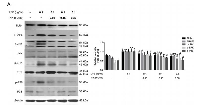
Application: WB Species: Mice Sample: RAW264.7 cells

Application: WB Species: Mouse Sample:
Restrictive clause
Affinity Biosciences tests all products strictly. Citations are provided as a resource for additional applications that have not been validated by Affinity Biosciences. Please choose the appropriate format for each application and consult Materials and Methods sections for additional details about the use of any product in these publications.
For Research Use Only.
Not for use in diagnostic or therapeutic procedures. Not for resale. Not for distribution without written consent. Affinity Biosciences will not be held responsible for patent infringement or other violations that may occur with the use of our products. Affinity Biosciences, Affinity Biosciences Logo and all other trademarks are the property of Affinity Biosciences LTD.