製品: MYOCD Antibody
カタログ: DF2434
タンパク質の説明: Rabbit polyclonal antibody to MYOCD
アプリケーション: WB IHC IF/ICC
Cited expt.: WB
反応性: Human, Mouse, Rat
予測: Bovine, Horse, Rabbit, Dog, Chicken, Xenopus
分子量: 100 kDa; 102kD(Calculated).
ユニプロット: Q8IZQ8
RRID: AB_2839640

類似製品を見る>>

   サイズ 価格 在庫状況
 100ul $280 在庫あり
 200ul $350 在庫あり

リードタイム: 当日配達

For pricing and ordering contact:
お問い合わせ先

製品説明

ソース:
Rabbit
アプリケーション:
WB 1:500-1:2000, IHC 1:50-1:200, IF/ICC 1:100-1:500
*The optimal dilutions should be determined by the end user. For optimal experimental results, antibody reuse is not recommended.
*Tips:

WB: For western blot detection of denatured protein samples. IHC: For immunohistochemical detection of paraffin sections (IHC-p) or frozen sections (IHC-f) of tissue samples. IF/ICC: For immunofluorescence detection of cell samples. ELISA(peptide): For ELISA detection of antigenic peptide.

反応性:
Human,Mouse,Rat
予測:
Bovine(89%), Horse(89%), Rabbit(100%), Dog(89%), Chicken(100%), Xenopus(88%)
クローナリティ:
Polyclonal
特異性:
MYOCD Antibody detects endogenous levels of total MYOCD.
RRID:
AB_2839640
引用形式: Affinity Biosciences Cat# DF2434, RRID:AB_2839640.
コンジュゲート:
Unconjugated.
精製:
The antiserum was purified by peptide affinity chromatography using SulfoLink™ Coupling Resin (Thermo Fisher Scientific).
保存:
Rabbit IgG in phosphate buffered saline, pH 7.4, 150mM NaCl, 0.02% sodium azide and 50% glycerol. Store at -20 °C. Stable for 12 months from date of receipt.
別名:

折りたたみ/展開

MYCD; MYCD_HUMAN; Myocardin; Myocd;

免疫原

免疫原:

A synthesized peptide derived from human MYOCD, corresponding to a region within the internal amino acids.

Uniprot:
遺伝子(ID):
発現特異性:
Q8IZQ8 MYCD_HUMAN:

Expressed in heart, aorta, and in smooth muscle cell-containing tissues: stomach, bladder, small intestine, colon, lung, placenta and uterus. Very faint expression in prostate and skeletal muscle.

タンパク質の説明:
Transcriptional factor that uses the canonical single or multiple CArG boxes DNA sequence. Binds CArG boxes only in the presence of serum response factor (SRF). Acts as a cofactor of SRF and modulates SRF-target genes. Regulates the expression of a set of cardiac and smooth muscle-specific genes. Plays a crucial role in cardiogenesis and differentiation of the smooth muscle cell lineage.
タンパク質配列:
MTLLGSEHSLLIRSKFRSVLQLRLQQRRTQEQLANQGIIPPLKRPAEFHEQRKHLDSDKAKNSLKRKARNRCNSADLVNMHILQASTAERSIPTAQMKLKRARLADDLNEKIALRPGPLELVEKNILPVDSAVKEAIKGNQVSFSKSTDAFAFEEDSSSDGLSPDQTRSEDPQNSAGSPPDAKASDTPSTGSLGTNQDLASGSENDRNDSASQPSHQSDAGKQGLGPPSTPIAVHAAVKSKSLGDSKNRHKKPKDPKPKVKKLKYHQYIPPDQKAEKSPPPMDSAYARLLQQQQLFLQLQILSQQQQQQQHRFSYLGMHQAQLKEPNEQMVRNPNSSSTPLSNTPLSPVKNSFSGQTGVSSFKPGPLPPNLDDLKVSELRQQLRIRGLPVSGTKTALMDRLRPFQDCSGNPVPNFGDITTVTFPVTPNTLPNYQSSSSTSALSNGFYHFGSTSSSPPISPASSDLSVAGSLPDTFNDASPSFGLHPSPVHVCTEESLMSSLNGGSVPSELDGLDSEKDKMLVEKQKVINELTWKLQQEQRQVEELRMQLQKQKRNNCSEKKPLPFLAASIKQEEAVSSCPFASQVPVKRQSSSSECHPPACEAAQLQPLGNAHCVESSDQTNVLSSTFLSPQCSPQHSPLGAVKSPQHISLPPSPNNPHFLPSSSGAQGEGHRVSSPISSQVCTAQMAGLHSSDKVGPKFSIPSPTFSKSSSAISEVTQPPSYEDAVKQQMTRSQQMDELLDVLIESGEMPADAREDHSCLQKVPKIPRSSRSPTAVLTKPSASFEQASSGSQIPFDPYATDSDEHLEVLLNSQSPLGKMSDVTLLKIGSEEPHFDGIMDGFSGKAAEDLFNAHEILPGPLSPMQTQFSPSSVDSNGLQLSFTESPWETMEWLDLTPPNSTPGFSALTTSSPSIFNIDFLDVTDLNLNSSMDLHLQQW

種類予測

種類予測:

Score>80(red) has high confidence and is suggested to be used for WB detection. *The prediction model is mainly based on the alignment of immunogen sequences, the results are for reference only, not as the basis of quality assurance.

Species
Results
Score
Rabbit
100
Chicken
100
Horse
89
Bovine
89
Dog
89
Xenopus
88
Pig
78
Sheep
78
Zebrafish
0
Model Confidence:
High(score>80) Medium(80>score>50) Low(score<50) No confidence

研究背景

機能:

Smooth muscle cells (SM) and cardiac muscle cells-specific transcriptional factor which uses the canonical single or multiple CArG boxes DNA sequence. Acts as a cofactor of serum response factor (SRF) with the potential to modulate SRF-target genes. Plays a crucial role in cardiogenesis and differentiation of the smooth muscle cell lineage (myogenesis) (By similarity).

PTMs:

Phosphorylation regulates negatively the intrinsic myocardin transcriptional activity.

細胞の位置付け:

Nucleus.

Extracellular region or secreted Cytosol Plasma membrane Cytoskeleton Lysosome Endosome Peroxisome ER Golgi apparatus Nucleus Mitochondrion Manual annotation Automatic computational assertionSubcellular location
組織特異性:

Expressed in heart, aorta, and in smooth muscle cell-containing tissues: stomach, bladder, small intestine, colon, lung, placenta and uterus. Very faint expression in prostate and skeletal muscle.

タンパク質ファミリー:

The C-terminal region contains a general transcription activation domain. The N-terminal region, comprising a basic and a Gln-rich domain, confers transcriptional potency and specificity by mediating association with the MADS box of SRF. The basic domain may be required for nuclear localization. The SAP domain is important for transactivation and ternary complex formation (By similarity).

参考文献

1). SREBP1-mediated lipogenesis promotes dedifferentiation and senescence of vascular smooth muscle cells through epigenetic remodeling. Nature communications, 2025 (PubMed: 41453868) [IF=16.6]

Application: WB    Species: human    Sample:

Fig. 4: SREBP1 directly promotes the senescence-like transformation of VSMC by activating lipogenesis. A Correlation between the expressions of SREBPs target genes and VSMC contractile genes (upper left panel), SREBPs target genes and aging genes (lower left panel), SREBP1 target genes and VSMC contractile genes (upper right panel), and SREBP1 target genes and aging genes (lower right panel) in the carotid artery from healthy and atherosclerosis patients (GSE100927). Correlation and P value (two-tailed) are based on Pearson’s correlation. B Heatmap depicting the average expression level of VSMC contractile genes, SREBP1 related genes, aging genes, and fibroblast genes of each VSMC or fibroblast subgroups in the carotid artery from atherosclerosis patients (single-cell transcriptomic, GSE253903). C Relative mRNA expression levels of genes involved in the maintain of VSMC contractile phenotype of MOVAS transfected with siCtrl, si-Srebf1#3, or si-Srebf2#3. Data are presented as mean ± SD. Statistical significance is calculated with a one-way ANOVA followed by Dunnett’s multiple comparisons test (n = 6 biological replicates). D Representative images of cell contraction assay of MOVAS transfected with siCtrl, si-Srebf1#3, or si-Srebf2#3. E Schematic diagram of the agonistic target of LXR-623. F Immunoblot analyses of precursor-SREBP1/2 (p-SREBP1/2), and nucleic-SREBP1/2 (n-SREBP1/2) of MOVAS after DMSO or LXR-623 treatment. Band quantification is normalized to the first lane. G GSEA based transcription factors (TFs) enrichment analysis for the activated TFs of three RNA-seq datasets (CON vs. HU, CTRL vs. SM, and DMSO vs. LXR-623) is shown above. Details of the co-activated TFs (3 in total) are listed below. SREBP1 is highlighted. H GSEA plots showing normalized enrichment scores (NES) and normalized P-value (calculated with an empirical phenotype-based permutation test) for SREBP1 target genes from RNA-Seq (DMSO vs. LXR-623). I The key genes, regulatory processes (black arrows), and heatmap analysis (middle panel) involved in cholesterol synthesis (left panel) and fatty acid synthesis (right panel) pathways. The Log2-transformed fold change (FC) values are presented in parentheses. J Lipid level in MOVAS after DMSO or LXR-623 treatment by BODIPY staining. K Representative transmission electron microscopy images of MOVAS treated with DMSO or LXR-623. Lipid droplets are indicated by red arrows. Scale bar = 2 μm (left panel), 1 μm (zoom-in panel). L Representative images of the cell contraction assay of MOVAS treated with DMSO or LXR-623. Quantifications of cell contraction diameters are shown in the lower panel. Data are presented as mean ± SD. Statistical significance is calculated with a two-tailed unpaired t test (n = 5 biological replicates). M Immunoblot analyses of MYOCD, α-SMA, and SM22α of MOVAS treated with DMSO or LXR-623. Band quantification is normalized to the first lane. The non-specific bands are marked by “*”. N GSEA plots showing normalized enrichment scores (NES) and normalized P-value (calculated with an empirical phenotype-based permutation test) for VSMC contraction from RNA-Seq (DMSO vs. LXR-623). O Representative FACS images of MOVAS treated with DMSO or LXR-623 by SPiDER-βGal staining. P Representative images of β-Gal staining of MOVAS treated with H2O2 (positive control), DMSO or LXR-623. Scale bar = 200 μm.

2). Colchicine reduces neointima formation and VSMC phenotype transition by modulating SRF-MYOCD activation and autophagy. Acta pharmacologica Sinica, 2025 (PubMed: 39663419) [IF=6.9]

3). MBNL1 regulates isoproterenol‐induced myocardial remodelling in vitro and in vivo. JOURNAL OF CELLULAR AND MOLECULAR MEDICINE, 2021 (PubMed: 33295096) [IF=5.3]

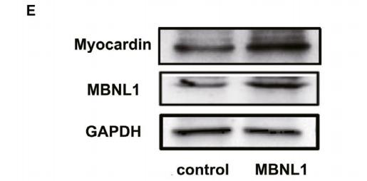
Application: WB    Species: mice    Sample: cardiomyocytes

FIGURE 3 MBNL1 increases Myocardin expression by binding to UGCU at the 3'-UTR of Myocardin mRNA. A, The 3'-UTR region of Myocardin contains the binding site of MBNL1. B, The binding of MBNL1 to Myocardin mRNA was validated using RIP. (n = 3, *, P < .05). C-F, The changes in Myocardin and MBNL1 in cardiomyocytes with overexpressed or silenced MBNL1 were measured using realtime PCR and Western blotting. (n = 3, *, P < .05, **, P < .01). G and H, Myocardin mRNA residues after 0, 20, 40 and 60 min in cardiomyocytes treated with Actinomycin D (ACD, 5 μg/mL) and with overexpressed or silenced MBNL1were measured using realtime PCR. I, The 3'-UTR region of Myocardin mRNA was transcribed in vitro and labelled with biotin. The combination of MBNL1 protein and 3'-UTR of Myocardin was detected using RNA pulldown assay. J, The RNA probes corresponding to nine prediction sites (probe # 10-18) and their corresponding antisense probes (probe # 1-9) were synthesized and biotin-labelled. The combination site of MBNL1 protein in the 3'-UTR of Myocardin was confirmed using RNA pull-down assay

4). LncRNA-Mhrt regulates cardiac hypertrophy by modulating the miR-145a-5p/KLF4/myocardin axis. JOURNAL OF MOLECULAR AND CELLULAR CARDIOLOGY, 2020 (PubMed: 31982428) [IF=4.9]

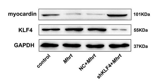
Application: WB    Species: mouse    Sample: primary cardiomyocytes

Fig. 2. |Mhrt regulates myocardin expression through KLF4 in primary cardiomyocytes.(D, E and F) Following knock down of endogenous KLF4 in primary cardiomyocytes, the effect of Mhrt on myocardin mRNA and protein levels was examined (n = 3, *,P < .05, **, P < .01, #, P > .05).

5). KLHL38 facilitates STS‐induced apoptosis in HL‐1 cells via myocardin degradation. IUBMB LIFE, 2022 (PubMed: 35112472) [IF=3.7]

Restrictive clause


Affinity Biosciences tests all products strictly. Citations are provided as a resource for additional applications that have not been validated by Affinity Biosciences. Please choose the appropriate format for each application and consult Materials and Methods sections for additional details about the use of any product in these publications.

For Research Use Only.
Not for use in diagnostic or therapeutic procedures. Not for resale. Not for distribution without written consent. Affinity Biosciences will not be held responsible for patent infringement or other violations that may occur with the use of our products. Affinity Biosciences, Affinity Biosciences Logo and all other trademarks are the property of Affinity Biosciences LTD.