GFP-Tag Rabbit Polyclonal Antibody - #T0006
| 製品: | GFP-Tag Rabbit Polyclonal Antibody |
| カタログ: | T0006 |
| タンパク質の説明: | Rabbit polyclonal antibody to GFP-Tag |
| アプリケーション: | WB IF/ICC |
| Cited expt.: | WB, IF/ICC |
| 反応性: | All |
| ユニプロット: | |
| RRID: | AB_2839423 |
製品説明
ソース:
Rabbit
アプリケーション:
WB 1:3000-1:10000, IF/ICC 1:100-1:500
*The optimal dilutions should be determined by the end user. For optimal experimental results, antibody reuse is not recommended.
*Tips:
*The optimal dilutions should be determined by the end user. For optimal experimental results, antibody reuse is not recommended.
*Tips:
WB: For western blot detection of denatured protein samples. IHC: For immunohistochemical detection of paraffin sections (IHC-p) or frozen sections (IHC-f) of tissue samples. IF/ICC: For immunofluorescence detection of cell samples. ELISA(peptide): For ELISA detection of antigenic peptide.
反応性:
All
クローナリティ:
Polyclonal
RRID:
AB_2839423
引用形式: Affinity Biosciences Cat# T0006, RRID:AB_2839423.
引用形式: Affinity Biosciences Cat# T0006, RRID:AB_2839423.
コンジュゲート:
Unconjugated.
精製:
The antiserum was purified by peptide affinity chromatography using SulfoLink™ Coupling Resin (Thermo Fisher Scientific).
保存:
Rabbit IgG in phosphate buffered saline, pH 7.4, 150mM NaCl, 0.02% sodium azide and 50% glycerol. Store at -20 °C. Stable for 12 months from date of receipt.
参考文献
1). Oryza sativa POSITIVE REGULATOR OF IRON DEFICIENCY RESPONSE 2 (OsPRI2) and OsPRI3 are involved in the maintenance of Fe homeostasis. PLANT CELL AND ENVIRONMENT, 2020
(PubMed: 31674679)
[IF=6.0]
Application: WB Species: plant Sample: plant cells
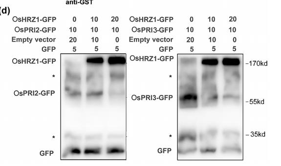
FIGURE 1| Interaction of OsHRZ1 with OsPRI2 and OsPRI3..(d) Degradation of OsPRI2 or OsPRI3 was carried out by detecting the OsPRI2/3‐GFP protein level in co‐infiltration experiments with increasing amounts of OsHRZ1‐GFP. GFP proteins were used as an internal control. Anti‐GFP antibody was used in western blot. Protein molecular weight (in kDa) is indicated. Stars indicate the non‐specific bands. Numbers indicate the ratio of the concentrations of agrobacteria used in co‐infiltration. Empty vector, a binary vector pOCA30 with a 35S promoter; GFP, 35S:GFP in pOCA30; OsPRI2‐GFP, 35S:OsPRI2‐GFP in pOCA30; OsPRI3‐GFP, 35S:OsPRI3‐GFP in pOCA30; OsHRZ1‐GFP, 35S:OsHRZ1‐GFP in pOCA30
Application: WB Species: Plant Sample: Root
FIGURE 1 Interaction of OsHRZ1 with OsPRI2 and OsPRI3. (a) Yeast two‐hybrid assays. Yeast co‐transformed with different BD and AD
plasmid combinations were spotted in parallel in 10‐fold dilution series on synthetic dropout medium lacking Leu/Trp/His/Ade. The C‐terminal
truncated OsHRZ1 and full‐length OsPRI2/3 were cloned into pGBKT7 and pGADT7, respectively. OsHRZ1‐C/OsPRI1, positive control. OsHRZ1‐
C/Empty, negative control. (b) Pull‐down assay. OsHRZ1 was fused with the GST tag and OsPRI2/3 were fused with the His tag. Recombinant
proteins were expressed in E. coli. Proteins were pulled down by glutathione Sepharose 4B and detected using the anti‐His antibody. Protein
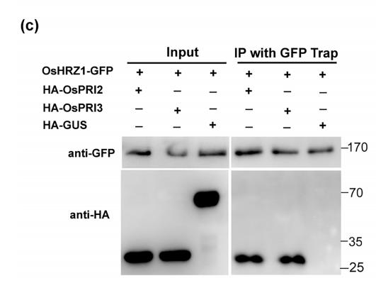
molecular weight (in kDa) is indicated. (c) CoIP assay. Total proteins from different combinations with OsHRZ1‐GFP and HA‐OsPRI2/HA‐OsPRI3/
HA‐GUS were immunoprecipitated with GFP‐Trap followed by immunoblotting with the indicated antibodies. HRZ1‐GFP/HA‐GUS, negative
control. Protein molecular weight (in kDa) is indicated. (d) Degradation of OsPRI2 or OsPRI3 was carried out by detecting the OsPRI2/3‐GFP
protein level in co‐infiltration experiments with increasing amounts of OsHRZ1‐GFP. GFP proteins were used as an internal control. Anti‐GFP
antibody was used in western blot. Protein molecular weight (in kDa) is indicated. Stars indicate the non‐specific bands. Numbers indicate the ratio
of the concentrations of agrobacteria used in co‐infiltration. Empty vector, a binary vector pOCA30 with a 35S promoter; GFP, 35S:GFP in
pOCA30; OsPRI2‐GFP, 35S:OsPRI2‐GFP in pOCA30; OsPRI3‐GFP, 35S:OsPRI3‐GFP in pOCA30; OsHRZ1‐GFP, 35S:OsHRZ1‐GFP in pOCA30.
(e) Cell‐free degradation. Ten‐day‐old roots grown in Fe‐sufficient solution were harvested and used for protein extraction. Incubation with or
without MG132 was performed over the indicated time course
2). TMT-based quantitative proteomics analysis reveals the attenuated replication mechanism of Newcastle disease virus caused by nuclear localization signal mutation in viral matrix protein. Virulence, 2020
(PubMed: 32420802)
[IF=5.5]
Application: WB Species: Mouse Sample: BSR-T7/5 cells
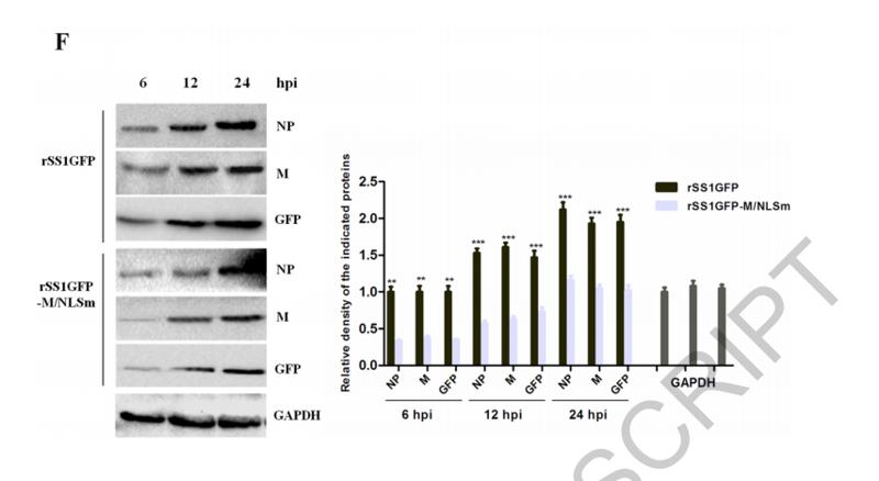
Figure 1. Nucleocytoplasmic trafficking of M protein promotes the replication and cytopathogenicity of NDV by affecting viral RNA synthesis and
transcription. (A) The subcellular localization of M protein in rSS1GFP- and rSS1GFP-M/NLSm-infected BSR-T7/5 cells at 6, 12, 18 and 24 hpi. DAPI was
used to stain nuclei. Original magnification was 1 × 200. (B) Virus titers were detected in BSR-T7/5 cells at the indicated time points. (C) The CPE and GFP
were observed in virus-infected BSR-T7/5 cells at 12 and 24 hpi. Original magnification was 1 × 200. (D) The viral RNA synthesis corresponding to the NP
and P genes and (E) viral transcription corresponding to the M and GFP genes in rSS1GFP- and rSS1GFP-M/NLSm-infected BSR-T7/5 cells were detected
by qRT-PCR. (F) The expression levels of NP, M and GFP proteins in rSS1GFP- and rSS1GFP-M/NLSm-infected BSR-T7/5 cells were examined by
Western blotting. The relative levels of the NP, M and GFP proteins were compared with the control GAPDH expression. Each data indicates the mean ± SD
of three independent experiments. P-values are indicated by asterisks (*P < 0.05, **P < 0.01, ***P < 0.001 compared to the value of rSS1GFP-M/NLSm).
3). Neuropilin-2 Signaling Modulates Mossy Fiber Sprouting by Regulating Axon Collateral Formation Through CRMP2 in a Rat Model of Epilepsy. MOLECULAR NEUROBIOLOGY, 2022
(PubMed: 36044155)
[IF=4.6]
4). Intracavernosal Adeno-Associated Virus-Mediated S100A1 Gene Transfer Enhances Erectile Function in Diabetic Rats by Promoting Cavernous Angiogenesis via VEGF-A/VEGFR2 Signaling. Journal of Sexual Medicine, 2019
(PubMed: 31378707)
[IF=3.3]
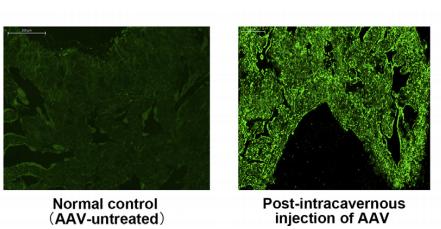
Application: IF/ICC Species: rat Sample: penis
Figure 2. |Transgene expression after intracavernosal AAV injection. Panel A shows the representative immunofluorescence images(magnification 100) of GFP protein at 4 weeks after intracavernosal delivery of AAV-GFP
Restrictive clause
Affinity Biosciences tests all products strictly. Citations are provided as a resource for additional applications that have not been validated by Affinity Biosciences. Please choose the appropriate format for each application and consult Materials and Methods sections for additional details about the use of any product in these publications.
For Research Use Only.
Not for use in diagnostic or therapeutic procedures. Not for resale. Not for distribution without written consent. Affinity Biosciences will not be held responsible for patent infringement or other violations that may occur with the use of our products. Affinity Biosciences, Affinity Biosciences Logo and all other trademarks are the property of Affinity Biosciences LTD.