製品: DRP1 Antibody
カタログ: DF7037
タンパク質の説明: Rabbit polyclonal antibody to DRP1
アプリケーション: WB IHC IF/ICC
Cited expt.: WB, IF/ICC
反応性: Human, Mouse, Rat
予測: Pig, Zebrafish, Bovine, Horse, Sheep, Rabbit, Dog, Chicken
分子量: 79kDa; 82kD(Calculated).
ユニプロット: O00429
RRID: AB_2838993

類似製品を見る>>

   サイズ 価格 在庫状況
 100ul $280 在庫あり
 200ul $350 在庫あり

リードタイム: 当日配達

For pricing and ordering contact:
お問い合わせ先

製品説明

ソース:
Rabbit
アプリケーション:
WB 1:500-1:2000, IHC 1:50-1:200, IF/ICC 1:100-1:500
*The optimal dilutions should be determined by the end user. For optimal experimental results, antibody reuse is not recommended.
*Tips:

WB: For western blot detection of denatured protein samples. IHC: For immunohistochemical detection of paraffin sections (IHC-p) or frozen sections (IHC-f) of tissue samples. IF/ICC: For immunofluorescence detection of cell samples. ELISA(peptide): For ELISA detection of antigenic peptide.

反応性:
Human,Mouse,Rat
予測:
Pig(100%), Zebrafish(83%), Bovine(100%), Horse(100%), Sheep(100%), Rabbit(100%), Dog(100%), Chicken(83%)
クローナリティ:
Polyclonal
特異性:
DRP1 Antibody detects endogenous levels of total DRP1.
RRID:
AB_2838993
引用形式: Affinity Biosciences Cat# DF7037, RRID:AB_2838993.
コンジュゲート:
Unconjugated.
精製:
The antiserum was purified by peptide affinity chromatography using SulfoLink™ Coupling Resin (Thermo Fisher Scientific).
保存:
Rabbit IgG in phosphate buffered saline , pH 7.4, 150mM NaCl, 0.02% sodium azide and 50% glycerol. Store at -20 °C. Stable for 12 months from date of receipt.
別名:

折りたたみ/展開

DLP1; dnm1l; DNM1L_HUMAN; Dnm1p/Vps1p-like protein; dnml1; DRP1; DVLP; Dymple; Dynamin 1 like; Dynamin family member proline-rich carboxyl-terminal domain less; Dynamin like protein; Dynamin related protein 1; Dynamin-1-like protein; Dynamin-like protein 4; Dynamin-like protein; Dynamin-like protein IV; Dynamin-related protein 1; DYNIV 11; EMPF; EMPF1; FLJ41912; HdynIV; VPS1;

免疫原

免疫原:

A synthesized peptide derived from human DRP1, corresponding to a region within C-terminal amino acids.

Uniprot:
遺伝子(ID):
発現特異性:
O00429 DNM1L_HUMAN:

Ubiquitously expressed with highest levels found in skeletal muscles, heart, kidney and brain. Isoform 1 is brain-specific. Isoform 2 and isoform 3 are predominantly expressed in testis and skeletal muscles respectively. Isoform 4 is weakly expressed in brain, heart and kidney. Isoform 5 is dominantly expressed in liver, heart and kidney. Isoform 6 is expressed in neurons.

タンパク質の説明:
Dynamin-related protein 1 (DRP1) is a member of the dynamin superfamily of GTPases. Members of this family have diverse cellular functions including vesicle scission, organelle fission, viral resistance, and intracellular trafficking (reviewed in 1). DRP1 affects mitochondrial morphology and is important in mitochondrial and peroxisomal fission in mammalian cells (2-5). The yeast ortholog of DRP1 clusters into a spiral-shaped structure on the mitochondrial membrane at the site of fission (reviewed in 6), and this structure is likely conserved in mammalian cells (3). The division of the mitochondria, which is required for apoptosis, as well as normal cell growth and development is controlled, in part, by the phosphorylation of DRP1 at Ser616 by Cdk1/cyclin B and at Ser637 by protein kinase A (PKA) (reviewed in 6). When phosphorylated at Ser616, DRP1 stimulates mitochondrial fission during mitosis. Conversely, fission is inhibited when DRP1 is phosphorylated at Ser637 (reviewed in 6). Dephosphorylation at Ser637 by calcineurin reverses this inhibition (7). In addition to phosphorylation, sumoylation of DRP1 is also an enhancer of mitochondrial fission (8). Balancing fission and fusion events is essential for proper mitochondrial function. Research studies have demonstrated mitochondrial defects in a variety of neurodegenerative diseases including Alzheimer’s disease, Parkinson’s disease, and Huntington’s disease (reviewed in 6).
タンパク質配列:
MEALIPVINKLQDVFNTVGADIIQLPQIVVVGTQSSGKSSVLESLVGRDLLPRGTGIVTRRPLILQLVHVSQEDKRKTTGEENGVEAEEWGKFLHTKNKLYTDFDEIRQEIENETERISGNNKGVSPEPIHLKIFSPNVVNLTLVDLPGMTKVPVGDQPKDIELQIRELILRFISNPNSIILAVTAANTDMATSEALKISREVDPDGRRTLAVITKLDLMDAGTDAMDVLMGRVIPVKLGIIGVVNRSQLDINNKKSVTDSIRDEYAFLQKKYPSLANRNGTKYLARTLNRLLMHHIRDCLPELKTRINVLAAQYQSLLNSYGEPVDDKSATLLQLITKFATEYCNTIEGTAKYIETSELCGGARICYIFHETFGRTLESVDPLGGLNTIDILTAIRNATGPRPALFVPEVSFELLVKRQIKRLEEPSLRCVELVHEEMQRIIQHCSNYSTQELLRFPKLHDAIVEVVTCLLRKRLPVTNEMVHNLVAIELAYINTKHPDFADACGLMNNNIEEQRRNRLARELPSAVSRDKSSKVPSALAPASQEPSPAASAEADGKLIQDSRRETKNVASGGGGVGDGVQEPTTGNWRGMLKTSKAEELLAEEKSKPIPIMPASPQKGHAVNLLDVPVPVARKLSAREQRDCEVIERLIKSYFLIVRKNIQDSVPKAVMHFLVNHVKDTLQSELVGQLYKSSLLDDLLTESEDMAQRRKEAADMLKALQGASQIIAEIRETHLW

種類予測

種類予測:

Score>80(red) has high confidence and is suggested to be used for WB detection. *The prediction model is mainly based on the alignment of immunogen sequences, the results are for reference only, not as the basis of quality assurance.

Species
Results
Score
Pig
100
Horse
100
Bovine
100
Sheep
100
Dog
100
Rabbit
100
Zebrafish
83
Chicken
83
Xenopus
0
Model Confidence:
High(score>80) Medium(80>score>50) Low(score<50) No confidence

研究背景

機能:

Functions in mitochondrial and peroxisomal division. Mediates membrane fission through oligomerization into membrane-associated tubular structures that wrap around the scission site to constrict and sever the mitochondrial membrane through a GTP hydrolysis-dependent mechanism. The specific recruitment at scission sites is mediated by membrane receptors like MFF, MIEF1 and MIEF2 for mitochondrial membranes. While the recruitment by the membrane receptors is GTP-dependent, the following hydrolysis of GTP induces the dissociation from the receptors and allows DNM1L filaments to curl into closed rings that are probably sufficient to sever a double membrane. Through its function in mitochondrial division, ensures the survival of at least some types of postmitotic neurons, including Purkinje cells, by suppressing oxidative damage. Required for normal brain development, including that of cerebellum. Facilitates developmentally regulated apoptosis during neural tube formation. Required for a normal rate of cytochrome c release and caspase activation during apoptosis; this requirement may depend upon the cell type and the physiological apoptotic cues. Plays an important role in mitochondrial fission during mitosis. Required for formation of endocytic vesicles. Proposed to regulate synaptic vesicle membrane dynamics through association with BCL2L1 isoform Bcl-X(L) which stimulates its GTPase activity in synaptic vesicles; the function may require its recruitment by MFF to clathrin-containing vesicles. Required for programmed necrosis execution. Rhythmic control of its activity following phosphorylation at Ser-637 is essential for the circadian control of mitochondrial ATP production.

Inhibits peroxisomal division when overexpressed.

Inhibits peroxisomal division when overexpressed.

PTMs:

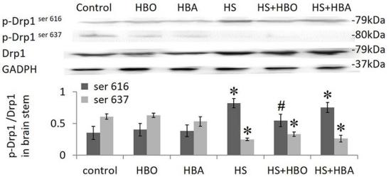
Phosphorylation/dephosphorylation events on two sites near the GED domain regulate mitochondrial fission. Phosphorylation on Ser-637 inhibits the GTPase activity, leading to a defect in mitochondrial fission promoting mitochondrial elongation. Dephosphorylated on this site by PPP3CA which promotes mitochondrial fission. Phosphorylation on Ser-616 activates the GTPase activity and promotes mitochondrial fission. Phosphorylated in a circadian manner at Ser-637.

Sumoylated on various lysine residues within the B domain, probably by MUL1. Sumoylation positively regulates mitochondrial fission. Desumoylated by SENP5 during G2/M transition of mitosis. Appears to be linked to its catalytic activity.

S-nitrosylation increases DNM1L dimerization, mitochondrial fission and causes neuronal damage.

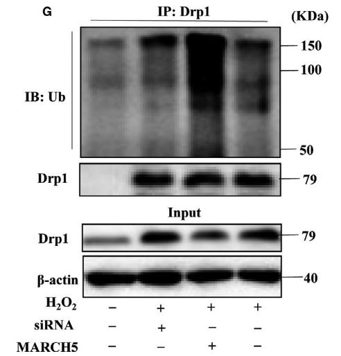
Ubiquitination by MARCHF5 affects mitochondrial morphology.

O-GlcNAcylation augments the level of the GTP-bound active form of DRP1 and induces translocation from the cytoplasm to mitochondria in cardiomyocytes. It also decreases phosphorylation at Ser-637 (By similarity).

細胞の位置付け:

Cytoplasm>Cytosol. Golgi apparatus. Endomembrane system>Peripheral membrane protein. Mitochondrion outer membrane>Peripheral membrane protein. Peroxisome. Membrane>Clathrin-coated pit. Cytoplasmic vesicle>Secretory vesicle>Synaptic vesicle membrane.
Note: Mainly cytosolic. Translocated to the mitochondrial membrane through O-GlcNAcylation and interaction with FIS1. Recruited to the mitochondrial outer membrane by interaction with MIEF1. Colocalized with MARCHF5 at mitochondrial membrane. Localizes to mitochondria at sites of division. Localizes to mitochondria following necrosis induction. Associated with peroxisomal membranes, partly recruited there by PEX11B. May also be associated with endoplasmic reticulum tubules and cytoplasmic vesicles and found to be perinuclear. In some cell types, localizes to the Golgi complex. Binds to phospholipid membranes.

Extracellular region or secreted Cytosol Plasma membrane Cytoskeleton Lysosome Endosome Peroxisome ER Golgi apparatus Nucleus Mitochondrion Manual annotation Automatic computational assertionSubcellular location
組織特異性:

Ubiquitously expressed with highest levels found in skeletal muscles, heart, kidney and brain. Isoform 1 is brain-specific. Isoform 2 and isoform 3 are predominantly expressed in testis and skeletal muscles respectively. Isoform 4 is weakly expressed in brain, heart and kidney. Isoform 5 is dominantly expressed in liver, heart and kidney. Isoform 6 is expressed in neurons.

タンパク質ファミリー:

The GED domain folds back to interact, in cis, with the GTP-binding domain and middle domain, and interacts, in trans, with the GED domains of other DNM1L molecules, and is thus critical for activating GTPase activity and for DNM1L dimerization.

Belongs to the TRAFAC class dynamin-like GTPase superfamily. Dynamin/Fzo/YdjA family.

研究領域

· Cellular Processes > Cell growth and death > Necroptosis.   (View pathway)

· Environmental Information Processing > Signal transduction > TNF signaling pathway.   (View pathway)

· Organismal Systems > Immune system > NOD-like receptor signaling pathway.   (View pathway)

参考文献

1). A thermo-sensitive hydrogel targeting macrophage reprogramming for sustained osteoarthritis pain relief. Acta pharmaceutica Sinica. B, 2025 (PubMed: 41311383) [IF=14.7]

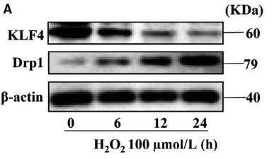
2). CircHIPK3 targets DRP1 to mediate hydrogen peroxide-induced necroptosis of vascular smooth muscle cells and atherosclerotic vulnerable plaque formation. Journal of advanced research, 2025 (PubMed: 38621622) [IF=11.4]

3). YAP1 inhibits the senescence of alveolar epithelial cells by targeting Prdx3 to alleviate pulmonary fibrosis. Experimental & molecular medicine, 2024 (PubMed: 38945958) [IF=9.5]

Application: WB    Species: Mouse    Sample: MLE-12 cell
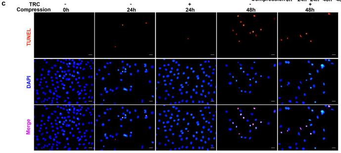
Fig. 6: Silencing of Prdx3 abolishes the effects of YAP1 on senescence and mitochondrial dysfunction. a Western blot analysis of p16 and p21 expression; n = 6. b SA-β-gal staining to evaluate senescence in MLE-12 cells transfected with the YAP1 overexpression plasmid or si-Prdx3 after BLM treatment; n = 6. Scale bars, 50 μm. The cell size was determined by confocal microscopy, and nuclei were stained with Hoechst 33342 (blue). Scale bars, 10 μm. c Western blot results showing the protein expression of Bax and Cyt-C in MLE-12 cells transfected with the YAP1 overexpression plasmid or si-Prdx3 after BLM treatment; n = 3. d Immunofluorescence staining of γH2AX in MLE-12 cells with YAP1 overexpression or Prdx3 silencing following BLM induction. Scale bars, 20 μm. e The protein levels of Drp1 and Mfn2 were measured by western blotting, n = 5. f Cytosolic and mitochondrial ROS levels in Prdx3-overexpressing MLE-12 cells after BLM administration were measured using a DCFH-DA probe and MitoSOX. Scale bars, 50 μm. g Representative images of MLE-12 cells stained with TMRM (red) and DAPI (nuclei; blue). Scale bars, 10 μm. h Representative images acquired by confocal microscopy showing morphological changes in mitochondria. Scale bars, 10 μm. i The O2 consumption rate was assessed using a Seahorse mitochondrial stress test. The data are presented as the means ± SEMs.

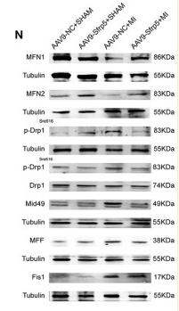
4). HSP70 attenuates compression-induced apoptosis of nucleus pulposus cells by suppressing mitochondrial fission via upregulating the expression of SIRT3. EXPERIMENTAL AND MOLECULAR MEDICINE, 2022 (PubMed: 35338257) [IF=9.5]

Application: IF/ICC    Species: Rat    Sample: NP cells
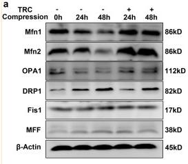
Fig. 2 HSP70 suppressed compression-induced apoptosis of NP cells. a, b The effects of TRC on apoptosis of NP cells measured by flow cytometry using Annexin V-FITC/PI staining. c Typical fluorescence photomicrograph of TUNEL staining of NP cells (scale bar: 50 μm). d, e The effects of VER on apoptosis of NP cells measured by flow cytometry using Annexin V-FITC/PI staining. f Typical fluorescence photomicrograph of TUNEL staining of NP cells (scale bar: 50 μm). (N = 3, **P < 0.01; ***P < 0.001).

Application: WB    Species: Rat    Sample: NP cells

Fig. 5 HSP70 suppressed compression-induced mitochondrial fission in NP cells. a The effects of TRC on the expression of DRP1, MFF, Fis1, Mfn1, Mfn2, and OPA1 (N = 3). b Representative fluorescence photomicrograph of TOM20 examined by immunofluorescence staining (N = 3, original magnification: ×1000). c The morphological ultrastructural appearance of mitochondria observed by TEM (N = 3). d The effects of VER on the expression of DRP1, MFF, Fis1, Mfn1, Mfn2, and OPA1 in NP cells treated with TRC (N = 3). e Representative fluorescence photomicrograph of TOM20 examined by immunofluorescence staining of NP cells treated with TRC and VER (N = 3, original magnification: ×1000). f Representative fluorescence photomicrograph of TOM20 examined by immunofluorescence staining of NP cells treated with TRC and HSP70-specific siRNAs (N = 3, original magnification: ×1000).

5). USP7 promotes cardiometabolic disorders and mitochondrial homeostasis dysfunction in diabetic mice via stabilizing PGC1β. Pharmacological research, 2024 (PubMed: 38815879) [IF=9.1]

Application: WB    Species: Mouse    Sample: heart

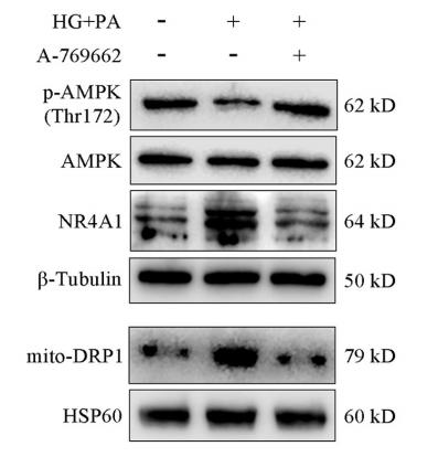
Fig. 6. USP7 inhibition improved cardiac mitochondrial dynamics in diabetic mice and NMCMs treated with HG+PA or H9c2 cells treated with PA. (A) Representative Western blot and quantification of Drp1, Fsi1, Mfn2 and Opa1 expression in the hearts of diabetic mice treated with P5091. n = 5 in each group. *P

6). Increased TSPO alleviates neuropathic pain by preventing pyroptosis via the AMPK-PGC-1α pathway. The journal of headache and pain, 2025 (PubMed: 39871133) [IF=7.3]

7). Zinc ions facilitate metabolic bioenergetic recovery post spinal cord injury by activating microglial mitophagy through the STAT3-FOXO3a-SOD2 pathway. Free radical biology & medicine, 2024 (PubMed: 39613048) [IF=7.1]

8). The Chinese herbal medicine Fufang Zhenzhu Tiaozhi ameliorates diabetic cardiomyopathy by regulating cardiac abnormal lipid metabolism and mitochondrial dynamics in diabetic mice. Biomedicine & pharmacotherapy = Biomedecine & pharmacotherapie, 2023 (PubMed: 37302318) [IF=6.9]

9). HSP70 alleviates sepsis-induced cardiomyopathy by attenuating mitochondrial dysfunction-initiated NLRP3 inflammasome-mediated pyroptosis in cardiomyocytes. Burns & trauma, 2022 (PubMed: 36439706) [IF=6.3]

Application: WB    Species: Rat    Sample: H9C2 cells
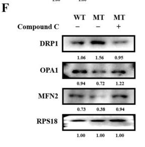
Figure 3.HSP70 improves sepsis-induced cardiomyopathy in vitro. H9C2 cells following LPS with/without HSP70 treatment to construct the septic model. (a) Cell morphology was observed and the amount of cell debris was calculated. (b) Cell viability measured by PI staining. Cell proliferation measured by (c) CCK8 and (d) crystal violet staining. (e) Levels of DRP1 determined by WB. Data are presented as the mean ± SD of data from three independent experiments (*p 

10). FGFR2 Mutation p.Cys342Arg Enhances Mitochondrial Metabolism-Mediated Osteogenesis via FGF/FGFR-AMPK-Erk1/2 Axis in Crouzon Syndrome. Cells, 2022 (PubMed: 36231091) [IF=6.0]

Application: WB    Species: Human    Sample:

Figure 7 Mitochondrial dynamics participate in osteogenesis mediated by FGFR/FGFR2-AMPK pathway: (A) Mito-tracker red and mitochondria 2D analysis showed more fragmented or punctate mitochondria with fewer and shorter branches in the MT group, while there were widely reticular mitochondria with longer branches in the WT group. (B) qRT-PCR and Western blot analysis demonstrated that the expressions of mitochondrial-fusion-related factors Mfn2 and Opa1 were downregulated, however, the expression of mitochondrial-fission-related factor Drp1 was upregulated in the MT group compared to the WT group. (C) Mito-tracker red and mitochondria 2D analysis showed a filamentous network of mitochondria with more and longer branches widely spread in the cytoplasm in the siFGFR2 group. (D) qPCR and Western blot analysis showed that the expression of fusion-related genes Mfn2 and Opa1 was increased and the expression of fission-related gene Drp1 was decreased in the siFGFR2 group. (E) After treatment with Compound C, the morphology of the mitochondria which should have tended to split adopted a fused reticular structure with more and longer branches, as shown by Mito-tracker red and mitochondria 2D analysis. (F) Western blot analysis showed the level of MFN2 and OPA1 was increased and then level of DRP1 was decreased, as induced by Compound C. p values were significant at * p < 0.05, ** p < 0.01, *** p < 0.001 and **** p < 0.0001.

もっと読みます

Restrictive clause


Affinity Biosciences tests all products strictly. Citations are provided as a resource for additional applications that have not been validated by Affinity Biosciences. Please choose the appropriate format for each application and consult Materials and Methods sections for additional details about the use of any product in these publications.

For Research Use Only.
Not for use in diagnostic or therapeutic procedures. Not for resale. Not for distribution without written consent. Affinity Biosciences will not be held responsible for patent infringement or other violations that may occur with the use of our products. Affinity Biosciences, Affinity Biosciences Logo and all other trademarks are the property of Affinity Biosciences LTD.