製品: Nrf2 Antibody
カタログ: AF0639
タンパク質の説明: Rabbit polyclonal antibody to Nrf2
アプリケーション: WB IHC IF/ICC
Cited expt.: WB, IHC, IF/ICC
反応性: Human, Mouse, Rat
予測: Pig, Bovine, Horse, Rabbit, Dog, Chicken
分子量: 100~120kD; 68kD(Calculated).
ユニプロット: Q16236
RRID: AB_2833793

類似製品を見る>>

   サイズ 価格 在庫状況
 100ul $280 在庫あり
 200ul $350 在庫あり

リードタイム: 当日配達

For pricing and ordering contact:
お問い合わせ先

製品説明

ソース:
Rabbit
アプリケーション:
WB 1:500-1:2000, IHC 1:50-1:200, IF/ICC 1:100-1:500
*The optimal dilutions should be determined by the end user. For optimal experimental results, antibody reuse is not recommended.
*Tips:

WB: For western blot detection of denatured protein samples. IHC: For immunohistochemical detection of paraffin sections (IHC-p) or frozen sections (IHC-f) of tissue samples. IF/ICC: For immunofluorescence detection of cell samples. ELISA(peptide): For ELISA detection of antigenic peptide.

反応性:
Human,Mouse,Rat
予測:
Pig(100%), Bovine(100%), Horse(88%), Rabbit(88%), Dog(100%), Chicken(100%)
クローナリティ:
Polyclonal
特異性:
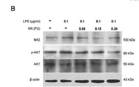
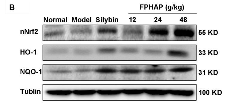
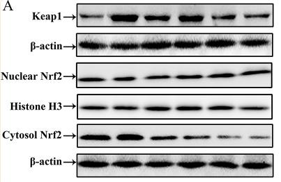
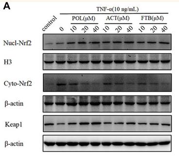
Nrf2 Antibody detects endogenous levels of total Nrf2.
RRID:
AB_2833793
引用形式: Affinity Biosciences Cat# AF0639, RRID:AB_2833793.
コンジュゲート:
Unconjugated.
精製:
The antiserum was purified by peptide affinity chromatography using SulfoLink™ Coupling Resin (Thermo Fisher Scientific).
保存:
Rabbit IgG in phosphate buffered saline , pH 7.4, 150mM NaCl, 0.02% sodium azide and 50% glycerol. Store at -20 °C. Stable for 12 months from date of receipt.
別名:

折りたたみ/展開

erythroid derived 2; HEBP1; like 2; NF E2 related factor 2; NF-E2-related factor 2; NF2L2_HUMAN; NFE2 related factor 2; NFE2-related factor 2; Nfe2l2; Nrf 2; NRF2; Nuclear factor (erythroid derived 2) like 2; Nuclear factor; nuclear factor erythroid 2 like 2; Nuclear factor erythroid 2 related factor 2; Nuclear factor erythroid 2-related factor 2; Nuclear factor erythroid derived 2 like 2;

免疫原

免疫原:

A synthesized peptide derived from human Nrf2, corresponding to a region within N-terminal amino acids.

Uniprot:
遺伝子(ID):
発現特異性:
Q16236 NF2L2_HUMAN:

Widely expressed. Highest expression in adult muscle, kidney, lung, liver and in fetal muscle.

タンパク質の説明:
GABPA Transcription factor capable of interacting with purine rich repeats (GA repeats). Necessary for the expression of the Adenovirus E4 gene. Belongs to the ETS family. Heterotetramer of two alpha and two beta subunits.
タンパク質配列:
MMDLELPPPGLPSQQDMDLIDILWRQDIDLGVSREVFDFSQRRKEYELEKQKKLEKERQEQLQKEQEKAFFAQLQLDEETGEFLPIQPAQHIQSETSGSANYSQVAHIPKSDALYFDDCMQLLAQTFPFVDDNEVSSATFQSLVPDIPGHIESPVFIATNQAQSPETSVAQVAPVDLDGMQQDIEQVWEELLSIPELQCLNIENDKLVETTMVPSPEAKLTEVDNYHFYSSIPSMEKEVGNCSPHFLNAFEDSFSSILSTEDPNQLTVNSLNSDATVNTDFGDEFYSAFIAEPSISNSMPSPATLSHSLSELLNGPIDVSDLSLCKAFNQNHPESTAEFNDSDSGISLNTSPSVASPEHSVESSSYGDTLLGLSDSEVEELDSAPGSVKQNGPKTPVHSSGDMVQPLSPSQGQSTHVHDAQCENTPEKELPVSPGHRKTPFTKDKHSSRLEAHLTRDELRAKALHIPFPVEKIINLPVVDFNEMMSKEQFNEAQLALIRDIRRRGKNKVAAQNCRKRKLENIVELEQDLDHLKDEKEKLLKEKGENDKSLHLLKKQLSTLYLEVFSMLRDEDGKPYSPSEYSLQQTRDGNVFLVPKSKKPDVKKN

種類予測

種類予測:

Score>80(red) has high confidence and is suggested to be used for WB detection. *The prediction model is mainly based on the alignment of immunogen sequences, the results are for reference only, not as the basis of quality assurance.

Species
Results
Score
Pig
100
Bovine
100
Dog
100
Chicken
100
Horse
88
Rabbit
88
Xenopus
75
Sheep
0
Zebrafish
0
Model Confidence:
High(score>80) Medium(80>score>50) Low(score<50) No confidence

研究背景

機能:

Transcription factor that plays a key role in the response to oxidative stress: binds to antioxidant response (ARE) elements present in the promoter region of many cytoprotective genes, such as phase 2 detoxifying enzymes, and promotes their expression, thereby neutralizing reactive electrophiles. In normal conditions, ubiquitinated and degraded in the cytoplasm by the BCR(KEAP1) complex. In response to oxidative stress, electrophile metabolites inhibit activity of the BCR(KEAP1) complex, promoting nuclear accumulation of NFE2L2/NRF2, heterodimerization with one of the small Maf proteins and binding to ARE elements of cytoprotective target genes. The NFE2L2/NRF2 pathway is also activated in response to selective autophagy: autophagy promotes interaction between KEAP1 and SQSTM1/p62 and subsequent inactivation of the BCR(KEAP1) complex, leading to NFE2L2/NRF2 nuclear accumulation and expression of cytoprotective genes. May also be involved in the transcriptional activation of genes of the beta-globin cluster by mediating enhancer activity of hypersensitive site 2 of the beta-globin locus control region.

PTMs:

Ubiquitinated in the cytoplasm by the BCR(KEAP1) E3 ubiquitin ligase complex leading to its degradation. In response to oxidative stress, electrophile metabolites, such as sulforaphane, modify KEAP1, leading to inhibit activity of the BCR(KEAP1) complex, promoting NFE2L2/NRF2 nuclear accumulation and activity. In response to autophagy, the BCR(KEAP1) complex is inactivated (By similarity).

Phosphorylation of Ser-40 by PKC in response to oxidative stress dissociates NFE2L2 from its cytoplasmic inhibitor KEAP1, promoting its translocation into the nucleus.

Acetylation at Lys-596 and Lys-599 increases nuclear localization whereas deacetylation by SIRT1 enhances cytoplasmic presence.

Glycation impairs transcription factor activity by preventing heterodimerization with small Maf proteins. Deglycation by FN3K restores activity.

細胞の位置付け:

Cytoplasm>Cytosol. Nucleus.
Note: Cytosolic under unstressed conditions: ubiquitinated and degraded by the BCR(KEAP1) E3 ubiquitin ligase complex (PubMed:15601839, PubMed:21196497). Translocates into the nucleus upon induction by electrophilic agents that inactivate the BCR(KEAP1) E3 ubiquitin ligase complex (PubMed:21196497).

Extracellular region or secreted Cytosol Plasma membrane Cytoskeleton Lysosome Endosome Peroxisome ER Golgi apparatus Nucleus Mitochondrion Manual annotation Automatic computational assertionSubcellular location
組織特異性:

Widely expressed. Highest expression in adult muscle, kidney, lung, liver and in fetal muscle.

タンパク質ファミリー:

The ETGE motif, and to a lower extent the DLG motif, mediate interaction with KEAP1.

Belongs to the bZIP family. CNC subfamily.

研究領域

· Genetic Information Processing > Folding, sorting and degradation > Protein processing in endoplasmic reticulum.   (View pathway)

· Human Diseases > Cancers: Overview > Pathways in cancer.   (View pathway)

· Human Diseases > Cancers: Specific types > Hepatocellular carcinoma.   (View pathway)

参考文献

1). Diamond-Like Carbon Depositing on the Surface of Polylactide Membrane for Prevention of Adhesion Formation During Tendon Repair. Nano-micro letters, 2024 (PubMed: 38687411) [IF=26.6]

Application: WB    Species: Rat    Sample:

Fig. 2 DLC suppress ROS production and oxidative stress both in vitro and in vivo. a Detection of cell ROS level on different membranes using ROS probes and b statistical analysis. c Diagram of animal experiment. d, f Content of MDA, 8-OHdG, and 3-NT in peritendinous tissues after 7 days of injury was examined by assay kit respectively. g Representative bands of Nrf2, HO-1, p-NF-κB were measured by Western blotting. h Nrf2 and HO-1 expression normalized to β-actin expression. i p-NF-κB expression normalized to NF-κB expression. Data represent independent experiments, and all data are presented as mean ± SD; NS non-significant,

2). Pyroptosis-responsive microspheres modulate the inflammatory microenvironment to retard osteoporosis in female mice. Nature communications, 2025 (PubMed: 40885754) [IF=16.6]

3). Morusin Alleviates Aortic Valve Calcification by Inhibiting Valve Interstitial Cell Senescence Through Ccnd1/Trim25/Nrf2 Axis. Advanced science (Weinheim, Baden-Wurttemberg, Germany), 2024 (PubMed: 38502885) [IF=15.1]

Application: WB    Species: human    Sample: VICs

Figure 5 Morusin alleviates the senescence of VICs through CCND1/Nrf2 pathway. A) VICs were transfected with CCND1 siRNA or scrambled siRNA, and then stimulated with OM for 7 days. Immunoblot analysis of Nrf2 expression in VICs from indicated groups (n = 3, each group). Bar plots showing the semiquantitative analysis of Nrf2 expression. B) Heatmap for NRF2, NQO1, and HMXO-1 in OM + morusin versus OM groups. C–E) Immunoblot analysis of NRF2 and HMXO-1 expression in VICs from indicated groups (n = 3, each group). Bar plots showing the fold change of indicated genes expression over control. F–H) Immunofluorescent staining of NRF2 (red), DCFH-DC (green), and DAPI (blue) in the VICs from indicated groups (n = 3, each group). Bar plots showing the semiquantitative analysis of fluorescence intensity. Scale bar 50 µm. I,J) Representative images showing MitoSOX (red) staining and quantification of the fluorescence intensity of MitoSOX fluorescence in VICs from indicated groups (n = 3, each group). Data are mean ± SD. *p < 0.05; **p < 0.01; ***p < 0.001 (ANOVA with Tukey's multiple comparisons test).

Application: IF/ICC    Species: human    Sample: VICs

Figure 5 Morusin alleviates the senescence of VICs through CCND1/Nrf2 pathway. A) VICs were transfected with CCND1 siRNA or scrambled siRNA, and then stimulated with OM for 7 days. Immunoblot analysis of Nrf2 expression in VICs from indicated groups (n = 3, each group). Bar plots showing the semiquantitative analysis of Nrf2 expression. B) Heatmap for NRF2, NQO1, and HMXO-1 in OM + morusin versus OM groups. C–E) Immunoblot analysis of NRF2 and HMXO-1 expression in VICs from indicated groups (n = 3, each group). Bar plots showing the fold change of indicated genes expression over control. F–H) Immunofluorescent staining of NRF2 (red), DCFH-DC (green), and DAPI (blue) in the VICs from indicated groups (n = 3, each group). Bar plots showing the semiquantitative analysis of fluorescence intensity. Scale bar 50 µm. I,J) Representative images showing MitoSOX (red) staining and quantification of the fluorescence intensity of MitoSOX fluorescence in VICs from indicated groups (n = 3, each group). Data are mean ± SD. *p < 0.05; **p < 0.01; ***p < 0.001 (ANOVA with Tukey's multiple comparisons test).

4). Piezo1 Upregulation in Monocyte-Derived Macrophages Impairs Post-Myocardial Infarction Cardiac Repair via Defective Efferocytosis and Enhanced Ferroptosis. Advanced science (Weinheim, Baden-Wurttemberg, Germany), 2025 (PubMed: 41214880) [IF=15.1]

Application: WB    Species: Mouse    Sample:

Figure 6 Piezo1 increased SLC7A11 in a Ca2+/ATF4-dependent manner and increased SLC15A3 in a Ca2+-dependent manner in macrophages. A) Heatmap of the genes of interest (the same as the 17 genes that were upregulated and the 17 genes that were downregulated by Yoda1 shown in Figure 5) identified by a new RNA-seq analysis of Piezo1fl/fl and Piezo1Lyz2 BMDMs, which were treated with DMSO or Yoda1 (5 µm) for 12 h. B) Levels of Atf4, Atf3, Nrf2 and P53 mRNAs in BMDMs after treatment with DMSO or Yoda1 (n = 6). C,D) Representative Western blots and quantification of the protein levels of ATF4, ATF3, NRF2 and P53 in BMDMs (n = 3). E) Representative images of immunofluorescence staining showing the ATF4 level and location in BMDMs treated with DMSO or Yoda1. F) Protein expression of ATF4 in the cytoplasm and nucleus of BMDMs treated with DMSO or Yoda1. G–I) Representative Western blots and quantification of ATF4 and SLC7A11 protein levels in BMDMs after control or Atf4 siRNA transfection and DMSO or Yoda1 treatment (n = 3). J) Flow cytometry analysis of intracellular Ca2+ levels by Fluo-4 staining in BMDMs from Piezo1fl/fl and Piezo1Lyz2 mice treated with DMSO or Yoda1 for 30 min (n = 5 mice per group). K) Fluo-4 fluorescence signals in BMDMs pretreated with 5 µM BAPTA-AM for 24 h followed by DMSO or 5 µm Yoda1 for 30 min were analyzed via flow cytometry (n = 3). L–O) Representative Western blots and quantification of the protein levels of ATF4, SLC7A11 and SLC15A3 in BMDMs (n = 3). The data in (B and D) were analyzed by unpaired Student's t test. Other data were analyzed by one-way ANOVA, followed by the Bonferroni post hoc correction.

5). Oral Metal-Free Melanin Nanozymes for Natural and Durable Targeted Treatment of Inflammatory Bowel Disease (IBD). Small (Weinheim an der Bergstrasse, Germany), 2023 (PubMed: 36760016) [IF=13.0]

6). Hydroxyl-augmented gold nanorods via lactone ring-opening alleviate cisplatin nephrotoxicity through Nrf2 activation. Biomaterials, 2026 (PubMed: 40763583) [IF=12.8]

7). Ultrasmall PtAu2 nanoclusters activate endogenous anti-inflammatory and anti-oxidative systems to prevent inflammatory osteolysis. Theranostics, 2023 (PubMed: 36793859) [IF=12.4]

8). Inhibition of macrophage inflammasome assembly and pyroptosis with GC-1 ameliorates acute lung injury. Theranostics, 2025 (PubMed: 39990234) [IF=12.4]

9). Mitochondria-targeted supramolecular coordination container encapsulated with exogenous itaconate for synergistic therapy of joint inflammation. Theranostics, 2023 (PubMed: 35547753) [IF=12.4]

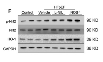
10). iNOS contributes to heart failure with preserved ejection fraction through mitochondrial dysfunction and Akt S-nitrosylation. Journal of Advanced Research, 2023 (PubMed: 36585107) [IF=11.4]

Application: WB    Species: Mouse    Sample: heart tissue

Fig. 6. iNOS inhibition alleviated oxidative stress in the heart of HFpEF mice. (A-C) Representative immunostaining and semi-quantification of NOX4 and SOD2 in heart tissue samples. (D) Myocardial malondialdehyde levels. (E) GSH-Px activity. (F-H) Representative immunostaining and semi-quantification of p-Nrf2/Nrf2 as well as HO-1 in the heart tissue samples from mice in different experimental groups. n = 6. The data are shown as mean ± SEM and were analyzed using one-way ANOVA followed by Tukey’s post hoc test. *, P < 0.05. **, P < 0.01. ***, P < 0.0005. ****, P < 0.0001. ns, no significant.

もっと読みます

Restrictive clause


Affinity Biosciences tests all products strictly. Citations are provided as a resource for additional applications that have not been validated by Affinity Biosciences. Please choose the appropriate format for each application and consult Materials and Methods sections for additional details about the use of any product in these publications.

For Research Use Only.
Not for use in diagnostic or therapeutic procedures. Not for resale. Not for distribution without written consent. Affinity Biosciences will not be held responsible for patent infringement or other violations that may occur with the use of our products. Affinity Biosciences, Affinity Biosciences Logo and all other trademarks are the property of Affinity Biosciences LTD.