.
Bands result from membrane strip incubation.")
Control Products

製品説明
*The optimal dilutions should be determined by the end user. For optimal experimental results, antibody reuse is not recommended.
*Tips:
WB: For western blot detection of denatured protein samples. IHC: For immunohistochemical detection of paraffin sections (IHC-p) or frozen sections (IHC-f) of tissue samples. IF/ICC: For immunofluorescence detection of cell samples. ELISA(peptide): For ELISA detection of antigenic peptide.
引用形式: Affinity Biosciences Cat# DF6487, RRID:AB_2838449.
折りたたみ/展開
Preproprotein S; Propiece of latent protein S; PROS 1; PROS; PROS_HUMAN; proS1; Protein S alpha; Protein Sa; PS 21; PS 22; PS 23; PS 24; PS 25; PS 26; PS21; PS22; PS23; PS24; PS25; PS26; PSA; THPH5; THPH6; Vitamin K dependent protein S; Vitamin K-dependent plasma protein S; Vitamin K-dependent protein S;
免疫原
A synthesized peptide derived from human PROS1, corresponding to a region within N-terminal amino acids.
- P07225 PROS_HUMAN:
- Protein BLAST With
- NCBI/
- ExPASy/
- Uniprot
MRVLGGRCGALLACLLLVLPVSEANFLSKQQASQVLVRKRRANSLLEETKQGNLERECIEELCNKEEAREVFENDPETDYFYPKYLVCLRSFQTGLFTAARQSTNAYPDLRSCVNAIPDQCSPLPCNEDGYMSCKDGKASFTCTCKPGWQGEKCEFDINECKDPSNINGGCSQICDNTPGSYHCSCKNGFVMLSNKKDCKDVDECSLKPSICGTAVCKNIPGDFECECPEGYRYNLKSKSCEDIDECSENMCAQLCVNYPGGYTCYCDGKKGFKLAQDQKSCEVVSVCLPLNLDTKYELLYLAEQFAGVVLYLKFRLPEISRFSAEFDFRTYDSEGVILYAESIDHSAWLLIALRGGKIEVQLKNEHTSKITTGGDVINNGLWNMVSVEELEHSISIKIAKEAVMDINKPGPLFKPENGLLETKVYFAGFPRKVESELIKPINPRLDGCIRSWNLMKQGASGIKEIIQEKQNKHCLVTVEKGSYYPGSGIAQFHIDYNNVSSAEGWHVNVTLNIRPSTGTGVMLALVSGNNTVPFAVSLVDSTSEKSQDILLSVENTVIYRIQALSLCSDQQSHLEFRVNRNNLELSTPLKIETISHEDLQRQLAVLDKAMKAKVATYLGGLPDVPFSATPVNAFYNGCMEVNINGVQLDLDEAISKHNDIRAHSCPSVWKKTKNS
種類予測
Score>80(red) has high confidence and is suggested to be used for WB detection. *The prediction model is mainly based on the alignment of immunogen sequences, the results are for reference only, not as the basis of quality assurance.
High(score>80) Medium(80>score>50) Low(score<50) No confidence
研究背景
Anticoagulant plasma protein; it is a cofactor to activated protein C in the degradation of coagulation factors Va and VIIIa. It helps to prevent coagulation and stimulating fibrinolysis.
The iron and 2-oxoglutarate dependent 3-hydroxylation of aspartate and asparagine is (R) stereospecific within EGF domains.
Secreted.
Plasma.
研究領域
· Organismal Systems > Immune system > Complement and coagulation cascades. (View pathway)
参考文献
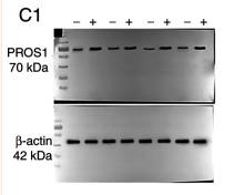
Application: WB Species: Human Sample: Wound granulation tissues
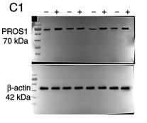
Application: WB Species: Human Sample: Wound granulation tissues
Restrictive clause
Affinity Biosciences tests all products strictly. Citations are provided as a resource for additional applications that have not been validated by Affinity Biosciences. Please choose the appropriate format for each application and consult Materials and Methods sections for additional details about the use of any product in these publications.
For Research Use Only.
Not for use in diagnostic or therapeutic procedures. Not for resale. Not for distribution without written consent. Affinity Biosciences will not be held responsible for patent infringement or other violations that may occur with the use of our products. Affinity Biosciences, Affinity Biosciences Logo and all other trademarks are the property of Affinity Biosciences LTD.