
, using PKM2 Antibody at 1/1000 dilution.
5ug/NC membrane strip.
Exposure for 30s with Affinity™ ECL Kit(#KF8001).
Bands result from membrane strip incubation.")


 and mouse anti-beta tubulin Ab(T0023 1:200) for 1 hour at 37°C. An AlexaFluor594 conjugated goat anti-rabbit IgG(H+L) Ab(Red) and an AlexaFluor488 conjugated goat anti-mouse IgG(H+L) Ab(Green) were used as the secondary antibody.
The nuclear counter stain is DAPI(blue).")
 promotes c-Myc protein degradation. Expression levels of c-Myc, HK2, PKM2, and LDHA were detected by Western blot (A) and qRT-PCR (B) analysis in MEG3 overexpression and knockdown colorectal cancer (CRC) cell lines. (C) MEG3 overexpression CRC cell lines were treated with 100 μg/ml of cycloheximide (CHX) and harvested at the indicated time points. c-Myc protein was detected by Western blot analysis, quantified by densitometry, and plotted against time to determine c-Myc stability. (D) CRC cells were transfected with pcDNA-c-Myc in combination with pcDNA-MEG3 in the presence of the HA-ubiquitin plasmid as indicated at the top. The cells were treated with MG132 (30 μM) for 6 h before harvesting, and the cell lysates were subjected to immunoprecipitation using anti-HA antibody. Ubiquitinated proteins were detected by Western blot with the anti-Flag antibody. (E) CRC cell lines that strongly express MEG3 were treated with 5 μM of MG132 for 12 h, and c-Myc protein was detected by Western blot. (F) Expression of FBXW7 was detected by Western blot in CRC cells that strongly and weakly expressed MEG3. (G) Level of FBXW7 was measured by Western blot in the pcDNA-MEG3 cells with MEG3 knockdown. Data are expressed as mean ± standard deviation from three independent experiments. *P < 0.05, **P < 0.01.")
 promotes c-Myc protein degradation. Expression levels of c-Myc, HK2, PKM2, and LDHA were detected by Western blot (A) and qRT-PCR (B) analysis in MEG3 overexpression and knockdown colorectal cancer (CRC) cell lines. (C) MEG3 overexpression CRC cell lines were treated with 100 μg/ml of cycloheximide (CHX) and harvested at the indicated time points. c-Myc protein was detected by Western blot analysis, quantified by densitometry, and plotted against time to determine c-Myc stability. (D) CRC cells were transfected with pcDNA-c-Myc in combination with pcDNA-MEG3 in the presence of the HA-ubiquitin plasmid as indicated at the top. The cells were treated with MG132 (30 μM) for 6 h before harvesting, and the cell lysates were subjected to immunoprecipitation using anti-HA antibody. Ubiquitinated proteins were detected by Western blot with the anti-Flag antibody. (E) CRC cell lines that strongly express MEG3 were treated with 5 μM of MG132 for 12 h, and c-Myc protein was detected by Western blot. (F) Expression of FBXW7 was detected by Western blot in CRC cells that strongly and weakly expressed MEG3. (G) Level of FBXW7 was measured by Western blot in the pcDNA-MEG3 cells with MEG3 knockdown. Data are expressed as mean ± standard deviation from three independent experiments. *P < 0.05, **P < 0.01.")
 The level of the consumption of glucose and the production of lactate was measured from the media of different groups. The fold change
between Cd-treated groups and non-treated control groups is represented using the Bar Chart (*P < .05 vs. cultivated 48 h non-treated control). (C) The relative
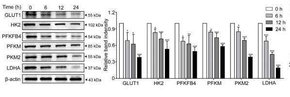
intracellular generation of ATP was measured (*P < .05 vs. Control). (D) Western blots of the relative aerobic glycolysis protein of GLUT1, HKII, PKM2 and LDHA in
both cells exposed to Cd for 0, 12, 24, 36 and 48 h. (E) Fold changes of them were normalized to the expression of β-actin. Each bar represents mean ± SD from three
independent experiments (*P < .05 vs. Control).")
 The intracellular activity levels of the metabolic enzymes HK2 and PKM2 were assessed by ELISA in Huh7, HepG2, and PLC/PRF/5 cells treated with 0.05 or 0.1 mg/mL ubenimex (**, P")
![PKM2 Antibody - Figure 5 BHD inhibited protein and mRNA expression of pyruvate kinase M2 (PKM2) and hypoxia-inducible factor-1 alpha (HIF-1α) as well as its target genes (GLUT1, PDK1, lactate dehydrogenase A [LDHA]) in aortas after high fat diet treatment.](http://img.affbiotech.cn/uploads/202409/5ce509e907ddfd09b9be49206226cfbb.png "Figure 5 BHD inhibited protein and mRNA expression of pyruvate kinase M2 (PKM2) and hypoxia-inducible factor-1 alpha (HIF-1α) as well as its target genes (GLUT1, PDK1, lactate dehydrogenase A [LDHA]) in aortas after high fat diet treatment. (a) Protein Levels of PKM2 were measured by Western blot analysis, and quantitative analysis was performed on the corresponding bands (n = 8–9). (b) Protein Levels of HIF-1α was measured by WB, and quantitative analysis was performed on the corresponding bands (n = 9). (c) The mRNA levels of GLUTA, (d) PDK1, and (e) LDHA in aortas (n = 6) were measured by qRT-PCR. Means ± standard error of mean. *P < 0.05, **P < 0.01, ***P < 0.001 versus Con; #P < 0.05, ##P < 0.01 versus Mod.")
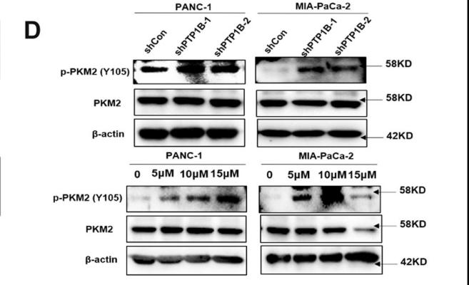
Western blot detected the expression levels of key autophagy-related proteins, including LC3B, P62 and Beclin1. GAPDH was defined as the internal control. (B)Quantitative statistics of protein expression levels of LC3B, P62 and Beclin1 after different treatments in BMSCs (n = 3). (C)Western blot detected the key protein expression levels of autophagic pathway, including p-mTOR, mTOR, p-ULK1, ULK1, PINK1 and Parkin1. GAPDH was defined as the internal control. (D)Quantitative statistics of protein expression levels of p-mTOR, mTOR, p-ULK1, ULK1, PINK1 and Parkin in different groups (n = 3). (E)Western blot detected the expression levels of PKM2 and p-PKM2 (Tyr105) in the three different groups. (F)Western blot detected the expression levels of p-AMPKα and total AMPKα after treatment of Shikonin in LVsh-PTP1B-transfected D-gal-induced Y-BMSCs. (G)Quantitative statistics of protein expression level of p-PKM2/PKM2 in the three different groups of D-gal-induced Y-BMSCs (n = 3). (H)Quantitative statistics of protein expression level of p-AMPKα/AMPKα after administration of Shikonin in LVsh-PTP1B-transfected D-gal-induced Y-BMSCs (n = 3). All data are shown as the mean ± SD. ****P ≤ 0.0001, ***P ≤ 0.001, **P < 0.01, *P < 0.05; NS, not significant (P > 0.05).")
.")

製品説明
*The optimal dilutions should be determined by the end user. For optimal experimental results, antibody reuse is not recommended.
*Tips:
WB: For western blot detection of denatured protein samples. IHC: For immunohistochemical detection of paraffin sections (IHC-p) or frozen sections (IHC-f) of tissue samples. IF/ICC: For immunofluorescence detection of cell samples. ELISA(peptide): For ELISA detection of antigenic peptide.
引用形式: Affinity Biosciences Cat# AF5234, RRID:AB_2837720.
折りたたみ/展開
CTHBP; Cytosolic thyroid hormone binding protein; Cytosolic thyroid hormone-binding protein; KPYM_HUMAN; MGC3932; OIP 3; OIP-3; OIP3; OPA interacting protein 3; Opa-interacting protein 3; p58; PK muscle type; PK, muscle type; PK2; PK3; PKM; PKM2; pykm; Pyruvate kinase 2/3; Pyruvate kinase 3; Pyruvate kinase isozymes M1/M2; Pyruvate kinase muscle; Pyruvate kinase muscle isozyme; pyruvate kinase PKM; Pyruvate kinase, muscle 2; TCB; THBP1; Thyroid hormone binding protein 1; Thyroid hormone binding protein cytosolic; Thyroid hormone-binding protein 1; Tumor M2 PK; Tumor M2-PK;
免疫原
A synthesized peptide derived from human PKM2, corresponding to a region within the internal amino acids.
Specifically expressed in proliferating cells, such as embryonic stem cells, embryonic carcinoma cells, as well as cancer cells.
- P14618 KPYM_HUMAN:
- Protein BLAST With
- NCBI/
- ExPASy/
- Uniprot
MSKPHSEAGTAFIQTQQLHAAMADTFLEHMCRLDIDSPPITARNTGIICTIGPASRSVETLKEMIKSGMNVARLNFSHGTHEYHAETIKNVRTATESFASDPILYRPVAVALDTKGPEIRTGLIKGSGTAEVELKKGATLKITLDNAYMEKCDENILWLDYKNICKVVEVGSKIYVDDGLISLQVKQKGADFLVTEVENGGSLGSKKGVNLPGAAVDLPAVSEKDIQDLKFGVEQDVDMVFASFIRKASDVHEVRKVLGEKGKNIKIISKIENHEGVRRFDEILEASDGIMVARGDLGIEIPAEKVFLAQKMMIGRCNRAGKPVICATQMLESMIKKPRPTRAEGSDVANAVLDGADCIMLSGETAKGDYPLEAVRMQHLIAREAEAAIYHLQLFEELRRLAPITSDPTEATAVGAVEASFKCCSGAIIVLTKSGRSAHQVARYRPRAPIIAVTRNPQTARQAHLYRGIFPVLCKDPVQEAWAEDVDLRVNFAMNVGKARGFFKKGDVVIVLTGWRPGSGFTNTMRVVPVP
種類予測
Score>80(red) has high confidence and is suggested to be used for WB detection. *The prediction model is mainly based on the alignment of immunogen sequences, the results are for reference only, not as the basis of quality assurance.
High(score>80) Medium(80>score>50) Low(score<50) No confidence
研究背景
Glycolytic enzyme that catalyzes the transfer of a phosphoryl group from phosphoenolpyruvate (PEP) to ADP, generating ATP. Stimulates POU5F1-mediated transcriptional activation. Plays a general role in caspase independent cell death of tumor cells. The ratio between the highly active tetrameric form and nearly inactive dimeric form determines whether glucose carbons are channeled to biosynthetic processes or used for glycolytic ATP production. The transition between the 2 forms contributes to the control of glycolysis and is important for tumor cell proliferation and survival. Promotes in a STAT1-dependent manner, the expression of the immune checkpoint protein CD274 in ARNTL/BMAL1-deficient macrophages (By similarity).
ISGylated.
Under hypoxia, hydroxylated by EGLN3.
Acetylation at Lys-305 is stimulated by high glucose concentration, it decreases enzyme activity and promotes its lysosomal-dependent degradation via chaperone-mediated autophagy.
FGFR1-dependent tyrosine phosphorylation is reduced by interaction with TRIM35.
Cytoplasm. Nucleus.
Note: Translocates to the nucleus in response to different apoptotic stimuli. Nuclear translocation is sufficient to induce cell death that is caspase independent, isoform-specific and independent of its enzymatic activity.
Specifically expressed in proliferating cells, such as embryonic stem cells, embryonic carcinoma cells, as well as cancer cells.
Belongs to the pyruvate kinase family.
研究領域
· Human Diseases > Endocrine and metabolic diseases > Type II diabetes mellitus.
· Human Diseases > Infectious diseases: Viral > Human papillomavirus infection.
· Human Diseases > Cancers: Overview > Viral carcinogenesis.
· Human Diseases > Cancers: Overview > Central carbon metabolism in cancer. (View pathway)
· Metabolism > Carbohydrate metabolism > Glycolysis / Gluconeogenesis.
· Metabolism > Nucleotide metabolism > Purine metabolism.
· Metabolism > Carbohydrate metabolism > Pyruvate metabolism.
· Metabolism > Global and overview maps > Metabolic pathways.
· Metabolism > Global and overview maps > Carbon metabolism.
· Metabolism > Global and overview maps > Biosynthesis of amino acids.
· Organismal Systems > Endocrine system > Glucagon signaling pathway.
参考文献
Application: WB Species: human Sample: MIA PaCa-2 cells
Application: WB Species: Human Sample: pancreatic cancer tissue

Application: WB Species: Mouse Sample: RAW 264.7 cells

Application: WB Species: Human Sample: MCF-7 cells

Application: WB Species: human Sample: LC cells
Restrictive clause
Affinity Biosciences tests all products strictly. Citations are provided as a resource for additional applications that have not been validated by Affinity Biosciences. Please choose the appropriate format for each application and consult Materials and Methods sections for additional details about the use of any product in these publications.
For Research Use Only.
Not for use in diagnostic or therapeutic procedures. Not for resale. Not for distribution without written consent. Affinity Biosciences will not be held responsible for patent infringement or other violations that may occur with the use of our products. Affinity Biosciences, Affinity Biosciences Logo and all other trademarks are the property of Affinity Biosciences LTD.