


 and mouse anti-beta tubulin Ab(T0023) for 1 hour at 37°C. An AlexaFluor594 conjugated goat anti-rabbit IgG(H+L) Ab(Red) and an AlexaFluor488 conjugated goat anti-mouse IgG(H+L) Ab(Green) were used as the secondary antibody.
The nuclear counter stain is DAPI(blue).")
 and mouse anti-beta tubulin Ab(#T0023) for 1 hour at 37°C. An AlexaFluor594 conjugated goat anti-rabbit IgG Ab(Red) and an AlexaFluor488 conjugated goat anti-mouse IgG Ab(Green) were used as the secondary antibody.
The nuclear counter stain is DAPI (blue).")
 Photographs of the PASMCs migration through the polycarbonate membrane stained by 0.2% crystal violet in hypoxia and treated with increasing concentrations of quercetin for 24 h. (B) Quantification of the number of cells migrating through the polycarbonate membrane of average of 3 independent experiments. (C) Full-length blots of MMP-2, MMP-9, CXCR4, Integrin α1, β1, and α5 and GAPDH are presented. (D) Results were quantified by densitometry analysis of the bands form (C) and then normalization to GAPDH protein. *Po0.05, **Po0.01 compared with control; #Po0.05, ##Po0.01 compared with hypoxia and quercetin treated PASMCs.")

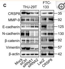
 microscopy at x200 magnification was used to assess cell morphology. The A549 cells (parental cells) had an epithelioid, rounded cobblestone appearance and there was limited formation of pseudopodia. A549/PTX and A549/DDP cells exhibited a spindle-shaped morphology and an increased formation of pseudopodia, indicating a loss of cell polarity. (B) E-cadherin, β-catenin, vimentin, MMP-2 and MMP-9 which are EMT-related proteins, were assessed in terms of expression levels. EMT-related transcription factors (Snail, Slug, Twist and ZEB1) were measured in A549/PTX and A549/DDP cells using western blot analysis. (C) The expression changes were confirmed at the mRNA level by qRT-PCR. Expression was standardized to the expression of GAPDH and normalized to 1.0 in the parental cells (compared with the parental A549 cells, means ± SEM, n=3, * P<0.05)")
 By human cancer pathway PCR array, ectopic expression of BCL2L10 up- or down-regulated several genes related to tumor proliferation, apoptosis, metastasis and angiogenesis. (B) Western blot was performed to confirm the downstream gene expression regulated by BCL2L10 in HepG2 cells. GAPDH was used as an internal control. (C) Schematic diagram of the molecular events for BCL2L10 function as a tumor suppressor through regulating cell cycle, proliferation, apoptosis metastasis and angiogenesis effectors.")
 MMP-9 protein expression. MMP-9 expression is expressed as the optical density ratio of MMP-9 to GAPDH.")
 was performed on lysates from the kidney cortex, and the band intensity ratios were quantified.")
 (upper panel) and in vitro (B) (lower panel);")
 Te expression of MMP2 and MMP9 at protein level was shown by Western blot. Band intensity is coming from densitometry, and data was shown as mean±SD")
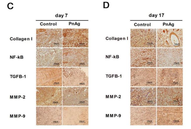
 Representative immunohistochemical staining of MMP2 and MMP9 in AFG1-induced lung adenocarcinoma.")
 Expression of MMP-7 and MMP-9 protein in A549 and SPC-A-1 cells after transfection.")


 Results of IHC assays. The expression levels of E-cadherin were significantly upregulated, whereas those of vimentin, MMP2, and MMP9 were downregulated by OA or regorafenib treatment, and OA enhanced the effects of regorafenib. The expression levels of iNOS and NT were upregulated by OA but not by regorafenib. (G) Staining indexes of IHC assays. Data are represented as mean ± standard error of the mean (*P < 0.05, **P < 0.01).")
 Apoptosis‐, proliferation‐ and migration‐related proteins were detected in HBEpC transfected with the NC or TRB3 plasmid. (d) Expression of p‐ERK and p‐JNK in HBEpC. Bcl2, B cell leukemia/lymphoma 2; ERK, extracellular signal‐regulated kinase; HBEpC, human bronchial epithelial cells; JNK, c‐Jun N‐terminalkinase; MMP9, matrix metallopeptidase 9;NC, negative control; TRB3, tribbles homolog 3")
 and western blot (down) were used to monitor the expression of EGFR signaling pathway and VM-related genes in 5–8F, and CNE1 after Foxq1 downregulation (B, C) or overexpression (D, E)")
 and western blot (H).")
 RT-qPCR
and western blot assays were used to detect expression changes of cellular functional genes and
proteins. (")
 RT-qPCR and western blot were used to
detect changes in TMEM92-AS1-related functional genes expression after knockdown of CCL5.
*P<0.05, **P<0.005 by Student’s t-test. For all of these experiments, values were mean±SD
(error bars) of triplicate samples in representative experiments.")

 The mRNA levels of MMP2, MMP7, MMP9 and MMP14 were detected in MCF7 and MDA-MB231 cells transfected with OE-MFAP5 or OE-Ctrl plasmids by qRT-PCR assay. (B) Western blot assay was used to investigate the expression of MMP2 and MMP9 in MCF7 and MDAMB-231 cells transfected with OE-MFAP5 plasmid or siRNAs or their controls. * means p < 0.05.")

 The bands of protein
expression. (B)-(F) Correlation analysis between the depression-like behaviors and protein expression. Statistical analyses of single comparison were conducted
through the Student’s t-test. All data are presented as mean ± SD, *p < 0.05, **p < 0.01, n = 3 per group.")
 Expression of proliferation- and migration-associated genes (PCNA, MMP9 and TIMP-1) were evaluated using western blotting in HEY cells. (C and D) Western blotting of proteins involved in integrin-β1-FAK signaling pathway in the KRT7-overexpressing HEY cells. (E) Expression of MMPs after knockdown of KRT7 in OVCAR433 cells. (F and G) Expression of the TGF-β signaling pathway-related proteins was evaluated by western blotting in KRT7-overexpressing HEY cells and KRT7-knockdown OVCAR433 cells. All experiments were performed at least three times. Results are presented as the mean ± standard deviation. **P<0.01. FAK, focal adhesion kinase; PCNA, proliferating cell nuclear antigen; FN, fibronectin; TIMP-1, TIMP metallopeptidase inhibitor 1; p-, phosphorylated; MMP, matrix metalloproteinase; KRT7, keratin 7; sh, short hairpin RNA; NC, negative control.")
 Transwell migration assay and transwell invasion assay were performed in LoVo cells with increasing concentration of recombinant GSN. (C) The expression of MMP2 and MMP9 were examined by western blot analysis in HT29 cell with increasing concentration of recombinant GSN.")
 The protein expression of SMARCE1, ERK1/2, p-ERK1/2, MMP9, and cyclin D1 was examined through western blotting. β-actin was used as control.")
 The Alizarin Red S (yellow arrow head) and TRAP staining (red arrow head) of bone tissues in groups. (B) Immunohistochemical analysis of bone tissue among groups for MMP9 and RUNX2 (x 400).")
 The Alizarin Red S (yellow arrow head) and TRAP staining (red arrow head) of bone tissues in groups. (B) Immunohistochemical analysis of bone tissue among groups for MMP9 and RUNX2 (x 400). (C, D) Western blotting results of MMP, RUNX2, Cath-K, OPG and RANKL expression in bone marrow from rat femurs. The data are expressed as the means ± SD (n = 6 in each group). ***P<0.001; ****P<0.0001 vs. SHAM, and #P<0.05; ####P<0.0001 vs. OVX by one-way ANOVA and Tukey’s post hoc test")

 NEAT1 expression; # indicates p < .01. (b) miR-204 expression; #indicates p < .01. (c) Western blot was to detect MMP-9 levels in serum exosomes.(d) MMP-9 expression; * indicates p < .05, # indicates p < .01")
 Western blot analysis of MMP-9 protein levels.")
. The change of cerebral edema rate in different groups (C). Representative protein (MMP-2and MMP-9) expression in different groups by
western blotting analysis (D–G). The date is expressed as the means ± S.E.M. (n = 3 rats in each group). *P < 0.05, **P < 0.01 and ***P < 0.001 vs MCAO
group.")
 of OS cells. (A) mRNA and (C, left column) protein levels of MMP2, MMP7, MMP9, N-cadherin, vimentin, E-cadherin and Snail after miR-708-5p overexpression (708-5p mimics) compared to the scramble negative control (NC mimic) in MG63 cells. (B) mRNA and (C, right column) protein levels of MMP2, MMP9, N-cadherin, vimentin, E-cadherin and Snail after miR-708-5p overexpression (708-5p mimics) compared to the scramble negative control (NC mimic) in SaOS-2 cells. Relative protein expression levels in (D) MG63 and (E) SaOS-2 cells. All data are presented as mean ± SD from at least three independent experiments. *P<0.05, **P<0.01, NC mimic vs. 708-5p mimic. OS, osteosarcoma; MMP, matrix metalloproteinase.")
 Representative western blotting images for MMP-2, MMP-9, and TNF-α in infrarenal aortas from three groups of animals; each lane represents one group. Bands intensities of pro-MMP-2 (B), active-MMP-2 (C), pro-MMP-9 (D), active-MMP-9 (E), and TNF-α (F) were normalized to corresponding GAPDH band and expressed as a fraction of control group, n = 5. *P < 0.05, **P < 0.01, ***P < 0.001, and ****P < 0.0001. Color images are available online.")
 Cell viability in the different transfection groups was eval‑uated by Cell Counting Kit‑8 assay. (B) Western blot analysis showing the protein expression levels of MMP2 and MMP9 in different groups.")
. *P < 0.05, vs. control group; #P < 0.05, vs. SE group (one-way analysis of variance followed by Newman-Keuls post hoc test). The experiment was repeated four times. AQP4: Aquaporin-4; HPA: heparanase; MMP-9: matrix metallopeptidase 9; SE: sepsis encephalopathy.")
 Representative immunofluorescence images and (B) quantification of MMP-9 in the ischemic brain of diabetic mice on day 7 post stroke. The expression of MMP-9 was significantly downregulated by CD28 SA administration on day 7 post stroke (n = 7–8). (C) Western blotting and (D) quantitative analysis showed that the
expression of MMP-9 was significantly decreased after CD28 treatment in diabetic mice on day 7 post stroke (n = 3–4). (E) The TBR values of NIRF
images were significantly correlated with the fluorescence integrated optical density (IOD) of MMP-9 on immunofluorescent staining, indicating the
high sensitivity of the probe for in vivo monitoring of the expression of MMP-9. Mean ± S.E.M. *p < 0.05.")
 Western blotting was used to assess MMP-9 and MMP-2 expression in cells treated with different doses of myricetin for 24 h.")
 The expression levels of metastasis-related proteins in MDA-MB-231 and BT-549 cells using western blotting analysis after treating with ADQ formula in a concentration-dependent manner.The results indicated that ADQ-formula treatment for 24 h dramatically attenuated the expression levels of proteins related to basement-membrane degradation and the EMT.")
 Cell viability in the different transfection groups was evaluated by Cell Counting Kit-8 assay. (B) Western blot analysis showing the protein expression levels of MMP2 and MMP9 in different groups. (C) Cell invasion and migration in different groups of cells after transfection. Scale bar, 100μm, ***P<0.001 vs. shRNA-NC; #P<0.05, ##P<0.01 and ###P<0.001 vs. shRNA-B3GALT5-AS1-1 + ov-NC. B3GALT5-AS1, β-1,3-galactosyltransferase 5-AS1; CSNK2A1, casein kinase 2 a1; MMP, matrix metalloproteinase; LncRNA, long non-coding RNA; Ov, expression; shRNA, short hairpin RNA; OD, optical density.")
 IHC staining was carried out to delineate the expression of PCNA protein in Skov3 cells. Real-time PCR was employed to detect the transcriptional levels of (B) P53 and (C) P21. (C, D, E, and F) Western blot analysis was conducted to measure key proteins of several signaling pathways. *, p<0.05 vs control group, * *, p<0.01 vs control group")
 Wound healing assay was employed to detect (A) HASMC and (B) A7R5 cell migration at 0 and 48 h after
incubation with different doses of myricetin (15, 30 and 60 μM) for 24 h. Scale bars, 200 μm. (C, D) Migration ratio was calculated as follows: ( wound width at 0 h −
wound width at 48 h)/wound width at 0 h × 100% (n = 3). (E) Transwell assay was used to detect the proliferative capability of VSMCs before incubation with
myricetin (15, 30 and 60 μM) for 24 h. Scale bars, 100 μm. (F, G) Quantification of the Transwell assay (n = 5). (H) Western blotting was used to assess MMP-9 and
MMP-2 expression in cells treated with different doses of myricetin for 24 h. (I-L) The densitometry analysis and quantitative results of (H) (n = 3). Data are displayed
as mean ± standard deviation (SD). * p < 0.05, ** p < 0.01 compared with the control group.")
 and RT-qPCR (b). c Effects of Vangl2 inhibition on MMP3, MMP9, MMP13, and IL-6 mRNA levels were measured by RT-qPCR. d Western blotting for MMP3, MMP9, MMP13, and IL-6 in three groups. Densitometric quantification values were normalized for GAPDH. **P < 0.01, ***P < 0.001")
 The HPLC fingerprints of the five independent
batches of ADQ formula samples as well as its major components including p-coumaric acid, calycosin-7-glucoside, liquiritin and glycyrrhizic acid and curcumol. The
HPLC fingerprints of five independent batches of ADQ formula were similar to one another. (B) The Effects of ADQ formula on cell proliferation in both the
nonmalignant mammary epithelial cells and the highly metastatic breast cancer cells. Colony formation assay for evaluating the long-term influence of ADQ formula
on MDA-MB-231 and BT-549 cells. The results showed that ADQ formula remarkably inhibited the proliferation of the breast cancer cell lines MDA-MB-231 and BT549 in a time- and dose-dependent manner, while having no obvious cytotoxicity in non-malignant cell lines including MCF-10A, HBL100, and HSF. (C-D) Wound
healing assay and transwell assay for evaluating the migration and invasion inhibitory effects of ADQ formula on MDA-MB-231 and BT-549 cells. The results
indicated that ADQ formula significantly inhibited cellular migratory and invasive capacities of MDA-MB-231 and BT-549 cells. (E) The expression levels of
metastasis-related proteins in MDA-MB-231 and BT-549 cells using western blotting analysis after treating with ADQ formula in a concentration-dependent manner.
The results indicated that ADQ-formula treatment for 24 h dramatically attenuated the expression levels of proteins related to basement-membrane degradation and
the EMT. All values are presented as the mean ± SD, n = 3, * p < 0.05, ** p < 0.01.")
 I-1 decreased MMP-9 production measured by immunostaining method. Magnification: 41.3 , Scale ¼ 50 mm. The blue fluorescence represents the nucleus and the green fluorescence represents the expression of MMP-9. (B)
Statistical data of MMP-9 from Western blot assay. (C) Statistical data of E-cadherin from Western blot assay. (D) Western blot images of MMP-9, E-cadherin and b-actin. Data are
presented as the mean ± SD of three independent assays. *P < 0.05; **P < 0.01, ***P < 0.001 compared with the C6 group. ###P < 0.001 compared with the sham group. TMZ
represents temozolomide.")
 and graph bar (B) showed changes in levels of MMP-9 immunoreactivity (green) in the trophoblast layer of placentas from all animal groups. Scale bar=50 μm. *P < 0.05 and **P < 0.01.")
. B Quantification of MYHC, MMP-9, TGF-β, and COL-1 expressions (n = 6). C-D Laser scanning confocal microscopy (LSCM) images show the levels of MYHC and COL-1 in H9c2 (n = 6; Scale bar represents as 50 μm). E-F The statistical analysis represents the positive expression of MYHC and COL-1 in H9c2. Data are represented as means ± SD, two-way ANOVA followed by Tukey’s post hoc test was used. *P < 0.05, **P < 0.01, and ***P < 0.001")
: representative immunoreactive bands of ODC, VEGF, MMP-9, GAPDH, total and phosphorylated Erk1/2 using specific antibody. (B–E): representative quantification of ODC, phosphorylated Erk1/2, VEGF, MMP-9. (F): determination of MMP-9 activity. * p < 0.05, ** p < 0.01, *** p < 0.001 compared with MCF-7/ADR cells, ## p < 0.01, ### p < 0.001, comparison between MCF-7/ADR group and MCF-7 group.")
 Apoptosis of the NPCs detected by flow cytometry analysis. (c, d) Typical fluorescence photomicrograph and quantitation of TUNEL staining of NPCs (scale bar: 100 μm). (e, f) Representative western blot bands and quantitation of the expression of cleaved-caspase 9, cleaved-caspase 3, Bax, and Bcl-2 in the NPCs. (g, h) Representative western blot bands and quantitation of the expression of anabolic mediators (aggrecan and collagen II) and catabolic mediators (MMP3 and MMP9). The data are represented as the mean ± SD from at least 3 independent experiments. ∗P < 0.05 and ∗∗P < 0.01 vs. the TBHP group.")
 The KEGG pathway enricherment analysis of DGEs. The length of the columns reflects the number of DGEs enrichment results are more significant. Different colours represent different P value. (B) Co-expression network map of differentially expressed genes KEGG enrichment. (C) Western blotting analysis of COL4A1, MMP2 and MMP9 expression in CNE1 and CNE2 cells. (D) Western blotting analysis of E-cadherin, N-cadherin, Vimentin and Snail expression in CNE1 and CNE2 cells. Data are shown as mean ± SD for three independent experiments, *p < 0.05, **p < 0.01 and ***p < 0.001.")
 After HSC-3 cells were treated with different concentrations (0, 0.625, 1.25, and 2.5 μM/L) of anlotinib for 24 h, expression of the p-Akt and Akt proteins was detected by immunofluorescence. (b) Quantitative immunofluorescence results of the p-Akt and Akt proteins. (c) HSC-3 cells were treated with anlotinib for 24 hours, and total MMP-2 and MMP-9, phosphorylated and total Akt and β-actin were detected by western blotting. (d) Ratio change of the migration markers MMP-2, MMP-9, phosphorylated, and total Akt. The data are expressed as the mean ± standard deviation of three repeats. ∗∗∗P < 0.001 compared with the control.")
 LAMC2 overexpression promotes integrin β1/FAK/Src/AKT protein expression in TU177 cells. (b) LAMC2 knockdown inhibited integrin β1/FAK/Src/AKT protein expression in AMC-HN-8 cells. Data were expressed as mean ± SD, n = 3. Compared to the vector group, #P < 0.05, ##P < 0.01. Compared to the shNC group, ∗P < 0.05, ∗∗P < 0.01.")
 and a transwell assay with matrigel for cell invasion (C) were performed on 786-O and ACHN cells following SSE treatment for 24 h. The images shown are representatives (Scale bar = 100 μm). Following SSE treatment for 6 h, E-cadherin, MMP-9 and VEGF were determined in cells using real-time RT PCR (D) and Western blot (E). β-Actin mRNA and β-actin were used as loading controls. The blots shown are representatives. The black bars and white bars denote 786-O cells and ACHN cells respectively. Data are presented as means ± SD (n ≥ 3). *P < 0.05 and **P < 0.01 vs the control group")
 The pathological changes in aorta tissues evaluated by H&E staining (scale bar = 500 μm). (b) Representative images of C/EBPα, PIK3C2A, LC3, MMP-2, and MMP-9 protein expression in aortic dissection rings determined by Western blot. (c) The expression level of indicated proteins")
 and (b). The BHLHE41, hypoxia-inducible factor-1alpha (HIF-1α), and epithelial-mesenchymal transition- (EMT-) related factor levels in hypoxia-induced CC cells were tested by Western blot. All experiments have been performed in triplicate, and data were expressed as mean ± SD. ∗∗P < 0.01 vs. hypoxia;")
 Western blot analysis was used to analyse the expression of G6PD in CRC cells transfected with the NC construct or shELFN1-AS1. (B) G6PD activity in the NC and shELFN1-AS1 groups. (C) Expression levels of G6PD protein in CRC cells with NC transfection, shELFN1-AS1 transfection or shELFN1-AS1+G6PD transfection ( ELFN1-AS1 knockout together with G6PD overexpression). (D) G6PD activity in CRC cells transfected with the NC construct, shELFN1-AS1 or shELFN1-AS1+G6PD. (E) A CCK-8 assay was used to evaluate proliferation in the different groups. (F) Transwell assays were used to detect the ability of transfected cells to migrate and invade. The histogram shows the numbers of migrating and invading cells (scale bar= 200 μm). (G) A 14-day colony formation experiment was conducted for the different groups. The histogram shows the number of cell colonies that formed. (H) Knockdown of ELFN1-AS1 in CRC cells enhanced the expression of E-cadherin and suppressed the expressions of N-cadherin and MMP-9, while overexpression of G6PD blocked these effects. Data are shown as the mean±SD.")
. C-F. The results of the Transwell invasion assay expressed as the number of invading cells in the experimental group compared to that in the respective control group (magnication: × 100, *P < 0.05, **P < 0.01, ***P < 1 0.001, Student's t test). G. circ-ITCH downregulated the mRNA expression of the cell migration-related genes MMP-2 and MMP-9 in PCa cells, as determined by qRT–PCR (*P < 0.05, **P < 0.01, Student’s t test). H. circ-ITCH downregulated the protein expression of MMP-2 and MMP-9 in PCa cells, as determined by Western blotting (*P < 0.05, **P < 0.01, Student’s t test).")
 The RT‐qPCR assay detected the relative mRNA expression of osteoclastogenesis-associated marker genes, including NFATc1, c-fos, TRAP, Rank, cathepsin K and MMP9. (B) Western-blot analysis for OB’s effects on protein levels of NFATc1 and c-fos. (C) Western-blot analysis for OB’s effects on protein levels of MMP9 and cathepsin K. (D) Quantitative analysis of NFATc1 and c-fos. (E) Quantitative analysis of MMP9 and cathepsin K. (F) Immunofluorescence images for OB’s effects on protein expression of NFATc1. ns, no statistical significance")
 The effect of Ecn on the migration of OS cells (Wound healing assay, 100 ×). (B) The effect of Ecn on the migration of OS cells (Transwell assay, 100 ×). (C) The effect of Ecn on the invasion of OS cells (Matrigel-coated Transwell assay, 100 ×). (D) The effect of Ecn on the protein level of MMP2, MMP7, MMP9, Snail, Vimentin, N-Cadherin and E-Cadherin in OS cells (Western blot). ##P")
, and the relative rates of migration were calculated (B). C and D, Transwell invasion assays of SW1736 and OCUT1 cells after treated with Mel or Da alone or their combination for 48 h. Representative images were shown (C), and the number of invading cells was calculated (D). E, The expression of EMT markers MMP‐9, E‐cadherin, N‐cadherin and vimentin was, respectively, detected in anaplastic thyroid cancer cells with indicated treatment by Western blot assays. Data were presented as mean ± SD of three independent experiments. The level of significance was indicated by ***P < .001, **P < .01")
 HTR-8/SVneo cell migration was inhibited upon transfection with miR-181b-5p mimics and enhanced upon transfection with miR-181b-5p inhibitor. (B) Transfected cells were assessed by Transwell assays. HTR-8/SVneo cell invasion was inhibited upon transfection with miR-181b-5p mimics and enhanced upon transfection with miR-181b-5p inhibitor. (C) Western blotting analysis showed that the expression levels of MMP-2 and MMP-9 were significantly decreased in miR-181b-5p mimics-treated cells and increased in miR-181b-5p inhibitor-treated cells compared with corresponding controls. The results are presented as the mean ± SD of at least three experimental repeats. *P")
 ANRIL inhibited proliferation and migration in platelet‐derived growth factor‐BB (PDGF‐BB)‐induced ASMCs through upregulating miR‐7‐5p. (A, B) The quantitative reverse transcriptase polymerase chain reaction (qRT‐PCR) is performed to measure the expression of lncRNA ANRIL and miR‐7‐5p in transfected cells; (C) Proliferation is tested by the MTT assay; (D, E) Migration is tested by the Transwell assay (bar = 50 μm); (F) western blot analysis is used to evaluate the expression levels of PCNA and MMP9; (G) PCNA/GAPDH and MMP9/GAPDH were determined; (H, I) qRT‐qPCR is used to evaluate the mRNA expression levels of PCNA and MMP9. n = 3; **p")
 Representative images and quantification of the wound healing assay. Scale bar, 200 µm. (B and C) The expression levels of MMP-2 and MMP-9 were determined using ELISA and western blotting. **P")
 The protein expression of CA9, IKBKB, NF-κB/p65, MMP-2, MMP-9, E-cadherin, and GAPDH in AGS cells was estimated by western blotting. (c, d) The protein expression of CA9, IKBKB, NF-κB/p65, MMP-2, MMP-9, E-cadherin, and GAPDH in MKN-45 cells was estimated by western blotting. (e, f) The mRNA expression of CA9, IKBKB, NF-κB/p65, MMP-2, MMP-9, and E-cadherin in AGS and MKN-45 cells treated with the control group, ArBu group (40 nM), CDDP group (40 μM), and ArBu combination with CDDP group (24.61 nM plus 28.37 μM). After treatment for 24 h, the mRNA level was detected by RT-PCR analysis. The data were presented as the mean (SD) of three independent experiments. *P < 0.05 and **P < 0.01, significantly different compared with the control treatment.")
 Functions and pathways negatively enriched for ImP treated cells and positively enriched for S1P treated cells were shown. The differentially expressed proteins was identified and analyzed by GO and KEGG pathway analysis. (C) Protein-protein interaction network analysis of ImP down-regulating protein by STRING. (D and E) The effects of S1P and ImP on the expression levels of VEGF, HIF-1α, CD31, and Ki67 proteins in HUVEC cells detected by Western blot. (F and G) The effect of ImP and S1P on the protein expression levels of MMP2, MMP9, Ki67 and Vimentin in NIH3T3cells under coculture conditions. (H) The effect of ImP on the cell membrane localization of RhoA. ImP-L:500 nM, ImP-H: 1μΜ. Data are expressed as the mean ± SD (∗p < 0.05, ∗∗p < 0.01). The number of sample replicates for all experiments was 3 (n = 3).")
 In vitro wound-healing experiments. (B) Relative protein expression of MMP9 and HPSE in vitro. (C) Immunofluorescence assay to determine the levels of MMP9 and HPSE in vitro. *P")
 In vitro wound-healing experiments. (B) Relative protein expression of MMP9 and HPSE in vitro. (C) Immunofluorescence assay to determine the levels of MMP9 and HPSE in vitro. *P")
 IL-1β, COX2, (C-D) IL-17, RORγt, and (E-F) MMP1, MMP3 and MMP9 were measured by western blot. Data are shown as the mean ± SD, #P")
 The effect of SEC on the migration of BC cells (Wound healing assay, 100 × ). (B) The effect of SEC on the migration of BC cells (Transwell assay, 100 × ). (C) The effect of SEC on the invasion of BC cells (Matrigel-coated transwell assay, 100 × ). (D) The effect of SEC on the protein levels of EMT markers and MMPs of BC cells (Western blot). Data are shown as mean ± SD from three independent experiments.")
 and Transwell assays (scale = 50 μm), respectively. 2 F: Changes in the levels of proteins related to the proliferation, apoptosis, and migration of HCT116 and SW480 cells after lncRNA RP11-197K6.1 knockdown, as analyzed by western blotting assay (**P")
. B. Western blotting analysis of the expression of VM-associated biomarkers after FGFR1 blockade. Representative images of the wound healing assays of SGC-7901 (C) and HGC-27 (D) cells after FGFR1 blockade (100 × magnification). E shows the fold changes in migration. Transwell assays showing the migration (F) and invasion (G) of GC cells after FGFR1 blockade (200 × magnification). The number of invading cells showing migration and invasion are shown in (H) and (I). WT: Wild Type; *p < 0.05, **p < 0.01.")
 After treatment with different concentrations of luteolin (0, 20, 40, 80, 160, 200 μM) for 48 h, the cell viabilities of RKO and SW480 cells were determined by using CCK-8 assay. (C, D) After treatment with 40 μM luteolin or DMSO (control) for 24 h, 48 h and 72 h, the cell proliferation capacities of RKO and SW480 cells were determined using CCK-8. (E, F) After treatment with 40 μM luteolin or DMSO (control) for 24 h, the cell proliferation capacities of RKO and SW480 cells were determined using EdU assay. Bar = 20 μm. (G, H) After treatment with 40 μM luteolin or DMSO (control) for 2 weeks, the cell colonies were stained with crystal violet to display the effect of luteolin on proliferation of RKO and SW480 cells. (I, J) After treatment with 40 μM luteolin or DMSO (control) for 72 h, the migration distance of RKO and SW480 cells were assayed by wounding healing experiments. (K, L) After treatment with 40 μM luteolin or DMSO (control) for 24h, the expression level of MMP3, MMP9, TIMP1 and VEGFA in RKO and SW480 cells was detected by qRT-PCR assay. (M) After treatment with 40 μM luteolin or DMSO (control) for 24h, the expression levels of MMP9, MMP3, PI3K, p-PI3K, AKT and p-AKT in RKO and SW480 cells was analyzed by Western blot. Data are presented as mean ± SD, * p < 0.05, ** p < 0.01, *** p < 0.001 compared with the control group.")
 VM formation ability was assessed in all of three types cells. (B and C ) The efficacy of PD-L1 knockdown using siRNA was evaluated through Western blotting and q-PCR. (D) The correlation between angiogenesis and PD-L1 levels was analyzed using the TCGA-Lung Adenocarcinoma (TCGA-LUAD) dataset, which comprised 465 samples. (E and F) VM-related genes’ expression in mRNA and protein levels was assessed through Western blotting and q-PCR analyses. (G) The number of VM structures decreased considerably following PD-L1 siRNA transfection")
 Effect of knockdown of KRT80 expression on migration properties of A549 cells. (B, D) Overexpression of KRT80 in H1299 cells accelerated cell migration. (E, F) The protein expression of MMP9, E-cadherin, Vimentin, N-cadherin in the KRT80 knockdown or overexpression cells were determined by western blot.")
 Western blotting was utilized to assess the protein expression levels of CD68, α-SMA, and MMP-9 in VSMCs following AIF-1 knockdown. (C, D) Western blotting was used to evaluate the protein expression levels of CD68, α-SMA, and MMP-9 in VSMCs that overexpress AIF-1. (E, G) Fluorescence photomicrographs display CD68 (red), α-SMA (green), and DAPI (blue) staining in VSMCs with AIF-1 knockdown. (F, H) Fluorescence photomicrographs illustrate the staining of CD68 (red), α-SMA (green), and DAPI (blue) in VSMCs that overexpress AIF-1. (I, J) Fluorescence photomicrographs show the staining of CD68 (red), α-SMA (green), and DAPI (blue) in atherosclerotic plaques of ApoE−/− mice fed a high-fat diet (HFD). Each experiment was conducted with three independent replicates. ∗P < 0.05, ∗∗P < 0.01, ∗∗∗P < 0.001. (For interpretation of the references to color in this figure legend, the reader is referred to the Web version of this article.)")
 and invasion (B) of lung cancer cells. CTC-TJH-01 and LLC cells were treated with the indicated concentrations (0, 0.5, 1 μmol·L−1) of TSZAF mc for 18 h, then transwell assays were used with and without Matrigel, images were taken by a microscope after staining with crystal violet. (C) The CTC-TJH-01 and LLC cells were exposed to TSZAF mc (0, 0.5, 1 μmol·L−1) for 24 h, the expression of MMP-2 and MMP-9 was detected by WB. β-Actin was used as an internal standard. Scale bar 100 μm. Each bar represents the mean ± SD of the three separate experiments. *P < 0.05, **P < 0.01, ***P < 0.001 vs the control group.")
 Cell proliferation was determined by CCK-8 assay after treatment with quercetin for 24 h, 48 h and 72 h. Cell invasion (B, C) and migration (D, E) was detected by transwell assay (scale bar = 100 μm). (F, G) Representative images and quantitative result of migration were detected by wound scratch assay (scale bar = 25 μm). (H) Expression of migration-related proteins E-cadherin, N-cadherin, MMP-9, MMP-2 and Snail in LOVO cells was determined by Western blot assay. ∗P < 0.05, ∗∗P < 0.01, ∗∗∗P < 0.001 vs. control. NC: Normoxia control; All other groups were treated in hypoxia.")
 IL-1β, IL-6, TNF-α and TGF-β1 expressions by western blot. (B) MMP2, MMP9 and MMP12 expressions by western blot. (C) Immunofluorescence staining for TGF-β1. (D) Immunofluorescence staining for MMP12. Data are presented as the mean ± SD. n = 3 per group.")

 Histograms of mRNA expression levels. (B) WB strip. (C) Protein expression histograms. *p < 0.05 vs. Control, #p < 0.05 vs. sh-HOXB9, ! p < 0.05 vs. sh-MMP12.")
 and epithelial-mesenchymal transition (EMT) signaling through CBX4. (A) We used Western blotting to measure the levels of MMP-associated proteins (MMP2 and MMP9) and EMT-associated proteins (E-Cadherin and N-Cadherin) in four groups of cells (HOS cells and MG-63 cells): shRNA-NC, shRNA-METTL3-1, shRNA-METTL3-1+OE-CBX4, and shRNA-METTL3-1+OE-NC. The differences in protein (MMP2, MMP9, E-cadherin, N-cadherin) expression in the four groups of cells were analyzed. The results are expressed as the mean ±SD of three independent experiments. (B) shows the calculated protein gray value. **p < 0.01, ***p < 0.001 using two-tailed Student’s t test.")
 Representative images of wound healing experiments and quantitative data of migration of HCC cells with upregulated or downregulated HOXB4 expression. Scale bar, 200 μm; (e-f) Invasion ability of HCC cells with upregulated or downregulated HOXB4 expression was determined by Transwell assays. Scale bar, 100 μm; (g) Immunoblots analysis of active MMP-2, active MMP-9, E-cadherin, N-cadherin, and Vimentin in Li-7 cells with upregulated or downregulated HOXB4 expression; (h) Representative immunofluorescence images of E-cadherin expression in Li-7 cells with upregulated or downregulated HOXB4 expression. Scale bar, 50 μm. Data are expressed as mean ± SD, N = 3. **p")
![MMP9 Antibody - Figure 11 Four hub genes (matrix metalloproteinase [MMP] 9, ATF3, CCL4, RELA) were upregulated in destabilization of the medial meniscus (DMM) mouse model.](http://img.affbiotech.cn/uploads/202512/55f2fd3a623ca7fccaca127aa2467fe8.png "Figure 11 Four hub genes (matrix metalloproteinase [MMP] 9, ATF3, CCL4, RELA) were upregulated in destabilization of the medial meniscus (DMM) mouse model. (A) Safranine O-Fast Green staining of cartilage in Sham and DMM group 4 weeks after surgery (100X and 400X). (B) The relative mRNA expression of MMP9, ATF3, CCL4, and RELA in mice cartilage tissues of each group. (C) The protein expression of MMP9, ATF3, CCL4, RELA/p65, and p-p65 in mice cartilage tissues of each group. (D) Quantification of relative protein expression by Western Blot.")
 Use CD55-carrying lentivirus to infect CT26 cells. Add 1 × 10⁴/well cells to a 96-well plate. Use the CCK8 method to measure the absorbance at 450 nm for 2 h, 4 h, 24 h, and 48 h. Use an empty virus as a control. (C) Repeat the above experiment using the Edu method. (D) Use CD55-carrying lentivirus to infect CT26 cells. Evenly place 200 cells in a small dish. Stain with Giemsa 2 weeks later and observe the results. (E) Use CD55-carrying lentivirus to infect CT26 cells. Add the cells to a small dish to ensure 5 × 10⁵/dish. On the second day, a horizontal line was drawn in each well with a pipette tip, and the cells were observed and photographed under a microscope 24 h later. (F) 1 × 10⁴/well was added to the upper chamber of the Transwell chamber, and crystal violet was used to stain and photographed 24 h later. (G, H) CT26 cells were infected with CD55 lentivirus, and the expression of EMT-related proteins was detected by Western Blot. GAPDH was used as an internal reference to analyze the relative gray value. The above experiments involved the sampling of quantitative data with mean ± SEM, and Student’s t-test was used for statistical analysis. At least three replicates were required for each group. P")
 The representative images of H&E staining. (B) The representative images of Masson’s staining. (n = 5) (C, D, E) The expression of MMP2 and MM9. (n = 3).")
 Results from proteomic analysis indicated that MDK affected the Wnt/β-catenin signalling pathway and protein ubiquitination. The raw data are shown in Table S8. (A) Volcano plot analysis results showed differentially expressed proteins (Table S9). The red dots represent upregulated expression in the siMDK group compared with the control group, and the yellow dots represent downregulated expression in the siMDK group compared with the control group. (B) Cluster analysis results showed similarities and differences among the groups (control vs. siMDK) and the stability of the mass spectrometry results of the three repeated experiments (Table S10). (C) KEGG pathway enrichment analysis revealed the signalling pathway affected by MDK knockdown (Table S11). (D) After treatment with 10 mM MG132 for 4 h, siMDK-U118MG and siMDK-SF126 cell lysates were immunoprecipitated with c-Myc and immunoblotted with ubiquitination antibodies. (E) After treatment with 10 mM MG132 for 4 h, OE-MDK-SHG44 and OE-MDK-U87 cell lysates were immunoprecipitated with c-Myc and immunoblotted with ubiquitination antibodies. (F–G) 50 mg/mL cycloheximide was added (at different time points: 0, 2, 4, 6, and 8 h) to glioma cells transfected with siRNA to block protein synthesis. The expression of c-Myc was then detected by Western blot. (H) MDK knockdown was performed on SF126, U118MG, and U251, and then a Western blot was used to detect the Wnt/β-catenin signalling pathway and EMT pathway markers. (I) MDK was overexpressed in U87, BT325, and SHG44, and Western blot was used to detect the Wnt/β-catenin signalling pathway and EMT pathway markers.")
 The mRNA expression levels of MMP9, MMP2 and MMP14 were detected by RT-PCR at P2 and P28.(n = 3 independent experiments, *P < 0.05,**P < 0.01). Data are presented as mean ± SD. (C) Immunostaining for MMP9 in Itgb4+/+and Itgb4−/−lungs. (n = 5,×200 magnification, scale bar, 50 μm).")
 qPCR results of AC16 cells with different treatment. (B) Heatmap of the previous qPCR results. (C–E) IF staining of Collagen I, TGF-β1 and MMP9 TGF-β1 of AC16 cells with different treatment. AC16 cells were preincubated with tFNAs (250 nM) or PBS for 12 h before being treated with DOX (1μM) for 12 h.")
 and MMP-9 protein (C) was detected by immunohistochemistry analysis. The representative samples labeled as a1-c1, a2-c2 and a3-c3 correspond to the parietal cortex, the CA2 region and the CA3 region, respectively. Bar = 50 μm. Mean optical density values of MMP-2 (B) and MMP-9 (D). Representative images of Western blot for MMP-2 protein and MMP-9 protein in brain (E). Quantitative analyses of MMP-2 (F) and MMP-9 (G) protein expression levels normalized to the internal control α-Tublin. The samples derive from the same experiment and that blots were processed in parallel. Original blots are presented in Supplementary Fig. 3. Data are shown as the mean ± SEM; n = 6 for each group. * P")
 ICT effect on the cytotoxicity of RAW 264.7 cells (n = 5). (B) Inhibition of OC differentiation by ICT was detected using TRAP staining (n = 5). (C) The effect of ICT on the expression of genes related to OC differentiation was assessed using RT-qPCR (n = 4). (D) MMP9 expression was detected following ICT treatment using western blot analysis (n = 4). (E) The effect of ICT on the expression levels of Esr1 and Esr2 was investigated using RTq-PCR (n = 4). (F) The effects of ICT on the protein levels of ESR1 and ESR2 were examined using western blot analysis (n = 4). *P < 0.05, **P < 0.01, ***P < 0.001 vs. the RANKL group, scale column:50 μm.")
 H&E staining showing cerebral oedema (scale: 20 µm). Black arrow: vacuolar changes of neuronal cells, red arrow: loose interstitial space, yellow arrow: Inflammatory cell infiltration. (b) Western blot bands for MMP-9, P-gp, MRP1, ZO-1 and Claudin5. (c) Relative protein level quantification (n=3/group; ANOVA with LSD-t test). (d) Sample source: ischaemic penumbra. Labels: I (ischaemic penumbra), II (infarction zone), III (contralateral brain). ANOVA: one way analysis of variance; BBB, blood-brain barrier; HDP, head-down position; MCAO/R, middle cerebral artery occlusion/reperfusion.")
 Representative IF micrographs of uterine sections from the implantation site on gestational days 5 and 6, stained with antibodies against HIF-2α (A) and RAB27B (B). Scale bar = 50 μm. (C and D) Analysis of relative fluorescence intensity of HIF-2α and RAB27B in the endometrial luminal epithelium at the implantation site on gestational day 5. The relative fluorescence intensity for each protein was measured and analysed using the Image J software. Each group contains 3 mice. Each point in the scatter plot represents the mean fluorescence intensity of the target protein in the luminal epithelium of a mouse, calculated from 5 random fields in the uterine section collected from the embryo implantation site in the endometrium of mice on gestational day 5. * indicates P")
 suppresses bone resorption activity of osteoclasts. a) Toluidine blue staining images of bovine bone slices in each group (100×, scale bar = 200 μm). b) and c) Expression and quantification of bone resorption-related proteins in osteoclasts of each group. d) Fluorescence expression of bone resorption-related proteins in each group (200×, scale bar = 100 μm). CTSK, cathepsin K; DAPI, 4′,6-diamidino-2-phenylindole; MMP-9, matrix metalloproteinase 9; TRAP, tartrate-resistant acid phosphatase.")
 RT-qPCR analysis of osteoclastogenic marker genes in BMMs treated with various concentrations of INS (0, 15, and 30 μM) for 5 days in the presence of RANKL and M-CSF. Relative mRNA expression levels of Acp5, c-Fos, Ctsk, and Nfatc1 were normalized to β-actin. (E, F) Representative Western blot images and quantification showing that INS dose-dependently upregulates the expression of antioxidant-related proteins (Nrf2, HO-1, SOD1, and catalase). (G, H) Representative Western blot images and quantification of osteoclastogenic marker proteins (CTSK, NFATc1, MMP9, and c-Fos) in RANKL-stimulated BMMs treated with INS (0, 15, and 30 μM) at days 1, 3, and 5 of differentiation. Data are presented as mean ± SD from three independent experiments performed in triplicate. *P < 0.05, **P < 0.01, ***P < 0.001 compared to the RANKL-stimulated control group (0 μM INS).")

製品説明
*The optimal dilutions should be determined by the end user. For optimal experimental results, antibody reuse is not recommended.
*Tips:
WB: For western blot detection of denatured protein samples. IHC: For immunohistochemical detection of paraffin sections (IHC-p) or frozen sections (IHC-f) of tissue samples. IF/ICC: For immunofluorescence detection of cell samples. ELISA(peptide): For ELISA detection of antigenic peptide.
引用形式: Affinity Biosciences Cat# AF5228, RRID:AB_2837714.
折りたたみ/展開
82 kDa matrix metalloproteinase-9; 92 kDa gelatinase; 92 kDa type IV collagenase; CLG 4B; CLG4B; Collagenase Type 4 beta; Collagenase type IV 92 KD; EC 3.4.24.35; Gelatinase 92 KD; Gelatinase B; Gelatinase beta; GelatinaseB; GELB; Macrophage gelatinase; MANDP2; Matrix metallopeptidase 9 (gelatinase B, 92kDa gelatinase, 92kDa type IV collagenase); Matrix Metalloproteinase 9; MMP 9; MMP-9; MMP9; MMP9_HUMAN; Type V collagenase;
免疫原
A synthesized peptide derived from human MMP9, corresponding to a region within C-terminal amino acids.
Detected in neutrophils (at protein level) (PubMed:7683678). Produced by normal alveolar macrophages and granulocytes.
- P14780 MMP9_HUMAN:
- Protein BLAST With
- NCBI/
- ExPASy/
- Uniprot
MSLWQPLVLVLLVLGCCFAAPRQRQSTLVLFPGDLRTNLTDRQLAEEYLYRYGYTRVAEMRGESKSLGPALLLLQKQLSLPETGELDSATLKAMRTPRCGVPDLGRFQTFEGDLKWHHHNITYWIQNYSEDLPRAVIDDAFARAFALWSAVTPLTFTRVYSRDADIVIQFGVAEHGDGYPFDGKDGLLAHAFPPGPGIQGDAHFDDDELWSLGKGVVVPTRFGNADGAACHFPFIFEGRSYSACTTDGRSDGLPWCSTTANYDTDDRFGFCPSERLYTQDGNADGKPCQFPFIFQGQSYSACTTDGRSDGYRWCATTANYDRDKLFGFCPTRADSTVMGGNSAGELCVFPFTFLGKEYSTCTSEGRGDGRLWCATTSNFDSDKKWGFCPDQGYSLFLVAAHEFGHALGLDHSSVPEALMYPMYRFTEGPPLHKDDVNGIRHLYGPRPEPEPRPPTTTTPQPTAPPTVCPTGPPTVHPSERPTAGPTGPPSAGPTGPPTAGPSTATTVPLSPVDDACNVNIFDAIAEIGNQLYLFKDGKYWRFSEGRGSRPQGPFLIADKWPALPRKLDSVFEERLSKKLFFFSGRQVWVYTGASVLGPRRLDKLGLGADVAQVTGALRSGRGKMLLFSGRRLWRFDVKAQMVDPRSASEVDRMFPGVPLDTHDVFQYREKAYFCQDRFYWRVSSRSELNQVDQVGYVTYDILQCPED
種類予測
Score>80(red) has high confidence and is suggested to be used for WB detection. *The prediction model is mainly based on the alignment of immunogen sequences, the results are for reference only, not as the basis of quality assurance.
High(score>80) Medium(80>score>50) Low(score<50) No confidence
研究背景
May play an essential role in local proteolysis of the extracellular matrix and in leukocyte migration. Could play a role in bone osteoclastic resorption. Cleaves KiSS1 at a Gly-|-Leu bond. Cleaves type IV and type V collagen into large C-terminal three quarter fragments and shorter N-terminal one quarter fragments. Degrades fibronectin but not laminin or Pz-peptide.
Processing of the precursor yields different active forms of 64, 67 and 82 kDa. Sequentially processing by MMP3 yields the 82 kDa matrix metalloproteinase-9.
N- and O-glycosylated.
Secreted>Extracellular space>Extracellular matrix.
Detected in neutrophils (at protein level). Produced by normal alveolar macrophages and granulocytes.
The conserved cysteine present in the cysteine-switch motif binds the catalytic zinc ion, thus inhibiting the enzyme. The dissociation of the cysteine from the zinc ion upon the activation-peptide release activates the enzyme.
Belongs to the peptidase M10A family.
研究領域
· Environmental Information Processing > Signal transduction > TNF signaling pathway. (View pathway)
· Human Diseases > Drug resistance: Antineoplastic > Endocrine resistance.
· Human Diseases > Infectious diseases: Viral > Hepatitis B.
· Human Diseases > Cancers: Overview > Pathways in cancer. (View pathway)
· Human Diseases > Cancers: Overview > Transcriptional misregulation in cancer.
· Human Diseases > Cancers: Overview > Proteoglycans in cancer.
· Human Diseases > Cancers: Overview > MicroRNAs in cancer.
· Human Diseases > Cancers: Specific types > Prostate cancer. (View pathway)
· Human Diseases > Cancers: Specific types > Bladder cancer. (View pathway)
· Organismal Systems > Immune system > IL-17 signaling pathway. (View pathway)
· Organismal Systems > Immune system > Leukocyte transendothelial migration. (View pathway)
· Organismal Systems > Endocrine system > Estrogen signaling pathway. (View pathway)
· Organismal Systems > Endocrine system > Relaxin signaling pathway.
参考文献

Application: WB Species: Human Sample: fibroblasts
Application: WB Species: Human Sample: thyroid cancer cells

Application: WB Species: human Sample:
Application: IHC Species: human Sample:

Application: WB Species: Mouse Sample: aortic tissues
Restrictive clause
Affinity Biosciences tests all products strictly. Citations are provided as a resource for additional applications that have not been validated by Affinity Biosciences. Please choose the appropriate format for each application and consult Materials and Methods sections for additional details about the use of any product in these publications.
For Research Use Only.
Not for use in diagnostic or therapeutic procedures. Not for resale. Not for distribution without written consent. Affinity Biosciences will not be held responsible for patent infringement or other violations that may occur with the use of our products. Affinity Biosciences, Affinity Biosciences Logo and all other trademarks are the property of Affinity Biosciences LTD.