, using GNAT1 Antibody at 1/1000 dilution.
5ug/NC membrane strip.
Exposure for 20s with Affinity™ ECL Kit(#KF8003).
Bands result from membrane strip incubation.")
 and mouse anti-beta tubulin Ab(T0023) for 1 hour at 37°C. An AlexaFluor594 conjugated goat anti-rabbit IgG(H+L) Ab(Red) and an AlexaFluor488 conjugated goat anti-mouse IgG(H+L) Ab(Green) were used as the secondary antibody.
The nuclear counter stain is DAPI(blue).")
| 製品: | GNAT1 Antibody |
| カタログ: | DF4109 |
| タンパク質の説明: | Rabbit polyclonal antibody to GNAT1 |
| アプリケーション: | WB IF/ICC |
| Cited expt.: | IF/ICC |
| 反応性: | Human, Mouse, Rat |
| 予測: | Zebrafish, Bovine, Horse, Sheep, Dog, Chicken, Xenopus |
| 分子量: | 36 KD(Observed); 40kD(Calculated). |
| ユニプロット: | P11488 |
| RRID: | AB_2836400 |
Control Products

製品説明
*The optimal dilutions should be determined by the end user. For optimal experimental results, antibody reuse is not recommended.
*Tips:
WB: For western blot detection of denatured protein samples. IHC: For immunohistochemical detection of paraffin sections (IHC-p) or frozen sections (IHC-f) of tissue samples. IF/ICC: For immunofluorescence detection of cell samples. ELISA(peptide): For ELISA detection of antigenic peptide.
引用形式: Affinity Biosciences Cat# DF4109, RRID:AB_2836400.
折りたたみ/展開
CSNBAD3; GBT1; GNAT1; GNAT1_HUMAN; GNATR; guanine nucleotide binding protein (G protein) alpha transducing activity polypeptide 1; guanine nucleotide binding protein G(T) alpha 1 subunit; Guanine nucleotide-binding protein G(t) subunit alpha-1; Rod specific transducin; rod-type transducin alpha subunit; Transducin alpha-1 chain; transducin, rod-specific;
免疫原
A synthesized peptide derived from human GNAT1, corresponding to a region within the internal amino acids.
Rod photoreceptor cells (PubMed:1614872). Predominantly expressed in the retina followed by the ciliary body, iris and retinal pigment epithelium (PubMed:22190596).
- P11488 GNAT1_HUMAN:
- Protein BLAST With
- NCBI/
- ExPASy/
- Uniprot
MGAGASAEEKHSRELEKKLKEDAEKDARTVKLLLLGAGESGKSTIVKQMKIIHQDGYSLEECLEFIAIIYGNTLQSILAIVRAMTTLNIQYGDSARQDDARKLMHMADTIEEGTMPKEMSDIIQRLWKDSGIQACFERASEYQLNDSAGYYLSDLERLVTPGYVPTEQDVLRSRVKTTGIIETQFSFKDLNFRMFDVGGQRSERKKWIHCFEGVTCIIFIAALSAYDMVLVEDDEVNRMHESLHLFNSICNHRYFATTSIVLFLNKKDVFFEKIKKAHLSICFPDYDGPNTYEDAGNYIKVQFLELNMRRDVKEIYSHMTCATDTQNVKFVFDAVTDIIIKENLKDCGLF
種類予測
Score>80(red) has high confidence and is suggested to be used for WB detection. *The prediction model is mainly based on the alignment of immunogen sequences, the results are for reference only, not as the basis of quality assurance.
High(score>80) Medium(80>score>50) Low(score<50) No confidence
研究背景
Functions as signal transducer for the rod photoreceptor RHO. Required for normal RHO-mediated light perception by the retina. Guanine nucleotide-binding proteins (G proteins) function as transducers downstream of G protein-coupled receptors (GPCRs), such as the photoreceptor RHO. The alpha chain contains the guanine nucleotide binding site and alternates between an active, GTP-bound state and an inactive, GDP-bound state. Activated RHO promotes GDP release and GTP binding. Signaling is mediated via downstream effector proteins, such as cGMP-phosphodiesterase (By similarity).
Cell projection>Cilium>Photoreceptor outer segment. Membrane>Peripheral membrane protein. Photoreceptor inner segment.
Note: Localizes mainly in the outer segment in the dark-adapted state, whereas is translocated to the inner part of the photoreceptors in the light-adapted state. During dark-adapted conditions, in the presence of UNC119 mislocalizes from the outer segment to the inner part of rod photoreceptors which leads to decreased photoreceptor damage caused by light.
Rod photoreceptor cells. Predominantly expressed in the retina followed by the ciliary body, iris and retinal pigment epithelium.
Belongs to the G-alpha family. G(i/o/t/z) subfamily.
研究領域
· Organismal Systems > Sensory system > Phototransduction.
参考文献
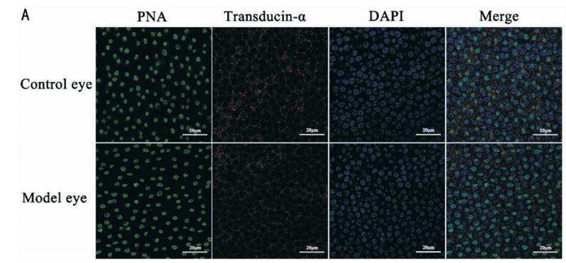
Application: IF/ICC Species: retina Sample: guinea pigs

Application: IF/ICC Species: Pig Sample: photoreceptor cells
Restrictive clause
Affinity Biosciences tests all products strictly. Citations are provided as a resource for additional applications that have not been validated by Affinity Biosciences. Please choose the appropriate format for each application and consult Materials and Methods sections for additional details about the use of any product in these publications.
For Research Use Only.
Not for use in diagnostic or therapeutic procedures. Not for resale. Not for distribution without written consent. Affinity Biosciences will not be held responsible for patent infringement or other violations that may occur with the use of our products. Affinity Biosciences, Affinity Biosciences Logo and all other trademarks are the property of Affinity Biosciences LTD.