製品: CLCN7 Antibody
カタログ: DF3932
タンパク質の説明: Rabbit polyclonal antibody to CLCN7
アプリケーション: WB IHC IF/ICC
Cited expt.: WB
反応性: Human, Mouse, Rat
予測: Bovine, Rabbit
分子量: 89 KD(Observed); 89kD(Calculated).
ユニプロット: P51798
RRID: AB_2836285

類似製品を見る>>

   サイズ 価格 在庫状況
 100ul $280 在庫あり
 200ul $350 在庫あり

リードタイム: 当日配達

For pricing and ordering contact:
お問い合わせ先

製品説明

ソース:
Rabbit
アプリケーション:
WB 1:500-1:1000, IF/ICC 1:100-1:500, IHC 1:50-1:200
*The optimal dilutions should be determined by the end user. For optimal experimental results, antibody reuse is not recommended.
*Tips:

WB: For western blot detection of denatured protein samples. IHC: For immunohistochemical detection of paraffin sections (IHC-p) or frozen sections (IHC-f) of tissue samples. IF/ICC: For immunofluorescence detection of cell samples. ELISA(peptide): For ELISA detection of antigenic peptide.

反応性:
Human, Mouse, Rat
予測:
Bovine(82%), Rabbit(%)
クローナリティ:
Polyclonal
特異性:
CLCN7 Antibody detects endogenous levels of total CLCN7.
RRID:
AB_2836285
引用形式: Affinity Biosciences Cat# DF3932, RRID:AB_2836285.
コンジュゲート:
Unconjugated.
精製:
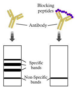
The antiserum was purified by peptide affinity chromatography using SulfoLink™ Coupling Resin.
保存:
Rabbit IgG in phosphate buffered saline , pH 7.4, 150mM NaCl, 0.02% sodium azide and 50% glycerol. Store at -20 °C. Stable for 12 months from date of receipt.
別名:

折りたたみ/展開

Chloride channel protein 7; CLC 7; ClC-7; ClC7; CLCN7; CLCN7_HUMAN; FLJ26686; FLJ39644; FLJ46423; H(+)/Cl(-) exchange transporter 7; OPTA2; OPTB4;

免疫原

免疫原:

A synthesized peptide derived from human CLCN7, corresponding to a region within N-terminal amino acids.

Uniprot:
遺伝子(ID):
発現特異性:
P51798 CLCN7_HUMAN:

Brain, testis, muscle and kidney.

タンパク質配列:
MANVSKKVSWSGRDRDDEEAAPLLRRTARPGGGTPLLNGAGPGAARQSPRSALFRVGHMSSVELDDELLDPDMDPPHPFPKEIPHNEKLLSLKYESLDYDNSENQLFLEEERRINHTAFRTVEIKRWVICALIGILTGLVACFIDIVVENLAGLKYRVIKGNIDKFTEKGGLSFSLLLWATLNAAFVLVGSVIVAFIEPVAAGSGIPQIKCFLNGVKIPHVVRLKTLVIKVSGVILSVVGGLAVGKEGPMIHSGSVIAAGISQGRSTSLKRDFKIFEYFRRDTEKRDFVSAGAAAGVSAAFGAPVGGVLFSLEEGASFWNQFLTWRIFFASMISTFTLNFVLSIYHGNMWDLSSPGLINFGRFDSEKMAYTIHEIPVFIAMGVVGGVLGAVFNALNYWLTMFRIRYIHRPCLQVIEAVLVAAVTATVAFVLIYSSRDCQPLQGGSMSYPLQLFCADGEYNSMAAAFFNTPEKSVVSLFHDPPGSYNPLTLGLFTLVYFFLACWTYGLTVSAGVFIPSLLIGAAWGRLFGISLSYLTGAAIWADPGKYALMGAAAQLGGIVRMTLSLTVIMMEATSNVTYGFPIMLVLMTAKIVGDVFIEGLYDMHIQLQSVPFLHWEAPVTSHSLTAREVMSTPVTCLRRREKVGVIVDVLSDTASNHNGFPVVEHADDTQPARLQGLILRSQLIVLLKHKVFVERSNLGLVQRRLRLKDFRDAYPRFPPIQSIHVSQDERECTMDLSEFMNPSPYTVPQEASLPRVFKLFRALGLRHLVVVDNRNQVVGLVTRKDLARYRLGKRGLEELSLAQT

種類予測

種類予測:

Score>80(red) has high confidence and is suggested to be used for WB detection. *The prediction model is mainly based on the alignment of immunogen sequences, the results are for reference only, not as the basis of quality assurance.

Species
Results
Score
Rabbit
100
Bovine
82
Pig
0
Horse
0
Sheep
0
Dog
0
Xenopus
0
Zebrafish
0
Chicken
0
Model Confidence:
High(score>80) Medium(80>score>50) Low(score<50) No confidence

研究背景

機能:

Slowly voltage-gated channel mediating the exchange of chloride ions against protons. Functions as antiporter and contributes to the acidification of the lysosome lumen.

細胞の位置付け:

Lysosome membrane>Multi-pass membrane protein.

Extracellular region or secreted Cytosol Plasma membrane Cytoskeleton Lysosome Endosome Peroxisome ER Golgi apparatus Nucleus Mitochondrion Manual annotation Automatic computational assertionSubcellular location
組織特異性:

Brain, testis, muscle and kidney.

タンパク質ファミリー:

Belongs to the chloride channel (TC 2.A.49) family. ClC-7/CLCN7 subfamily.

参考文献
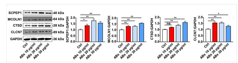
1). Donor MSCs release apoptotic bodies to improve myocardial infarction via autophagy regulation in recipient cells. Autophagy, 2020 (PubMed: 31959090) [IF=14.6]

Application: WB    Species: human    Sample: human umbilical vein endothelial cells (HUVECs)
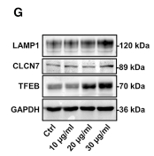
Figure S3. ABs upregulated the expression of lysosome-related proteins. The expression levels of SCPEP1, MCOLN1, CTSD and CLCN7 were detected by western blots in HUVECs treated with ABs (20 µg/ml). Quantitative analysis of the western blots was performed by ImageJ software. The results were representative of the data generated in at least 3 independent experiments and presented as mean ± SEM. ns, not significant; *P<0.05; **P<0.01 by one-way ANOVA.

2). Apoptotic vesicles activate autophagy in recipient cells to induce angiogenesis and dental pulp regeneration. Molecular Therapy, 2022 (PubMed: 35538661) [IF=12.1]

Application: WB    Species: Human    Sample: endothelial cells
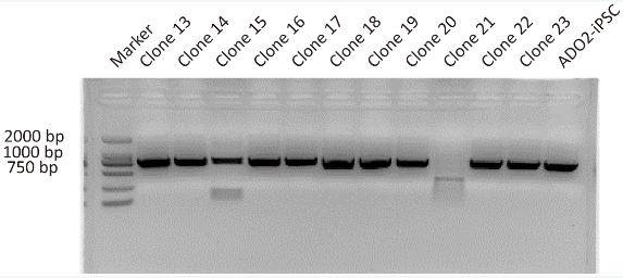
Figure 6 TUFM derived from hDPSC-apoVs activate EC autophagy and promote EC angiogenesis via the TFEB-induced autophagy-lysosome pathway (A) Western blotting analysis shows the presence of TUFM in apoVs derived from hDPSCs, si-NC-treated hDPSCs, and si-TUFM-treated hDPSCs. (B) Western blot analysis shows autophagy-associated gene (ATG7, Beclin-1, and LC3) expressions in ECs after incubation with apoVs or si-TUFM-apoVs. The angiogenic capacity of ECs, including tube formation (C), migration (D), and proliferation (E) were detected after treatment with apoVs or si-TUFM-apoVs. Scale bar, 100 μm. (F) Representative confocal microscopy images show the number of lysosomes in ECs after treatment with apoVs or Torin 1. Scale bar, 50 μm. (G) Western blot analysis shows lysosome-associated gene (LAMP1 and CLCN7) and TFEB expressions in ECs after incubation with apoVs at concentrations of 10, 20, or 30 μg/mL. (H) Expression of TFEB in cytosolic (Cyt.) and nuclear (Nuc.) fractions was detected by western blots after treatment with apoVs or Torin 1. (I) Representative confocal microscopy images show the locations of TFEB in ECs after treatment with apoVs or Torin1. Scale bar, 25 μm. (J) Western blot analysis shows autophagy-associated gene (ATG7, Beclin-1, and LC3) expressions in ECs after decreased expression of TFEB. (K) Western blot analysis shows autophagy-associated gene (ATG7, Beclin-1, and LC3) expressions in ECs after increased expression of TFEB. After transfection with si-TFEB, the angiogenic capacity of ECs, including tube formation (L), migration (M), and proliferation (N) were detected following coculture with apoVs (20 μg/mL). Scale bar, 100 μm; n = 3-5 per group. Data are presented as mean ± SD. Statistical analyses were performed by Student’s t test (two-tailed) for two-group comparisons and one-way ANOVA with Tukey’s post hoc test or Welch’s ANOVA with Games-Howell post hoc test for multiple-group comparisons.

3). CRISPR/Cas9-Mediated Gene Correction in Osteopetrosis Patient-Derived iPSCs. Frontiers in Bioscience-Landmark, 2023 (PubMed: 37395026) [IF=3.3]

Application: WB    Species: Mouse    Sample:

Fig. 2. Representative images showing the PCR assay for CLCN7 detection in different GC-ADO2-iPSC clones and ADO2-iPSCs. Clone 13-clone23 represent GC-ADO2-iPSC clone 13- GC-ADO2-iPSC clone23.

Restrictive clause


Affinity Biosciences tests all products strictly. Citations are provided as a resource for additional applications that have not been validated by Affinity Biosciences. Please choose the appropriate format for each application and consult Materials and Methods sections for additional details about the use of any product in these publications.

For Research Use Only.
Not for use in diagnostic or therapeutic procedures. Not for resale. Not for distribution without written consent. Affinity Biosciences will not be held responsible for patent infringement or other violations that may occur with the use of our products. Affinity Biosciences, Affinity Biosciences Logo and all other trademarks are the property of Affinity Biosciences LTD.