
, using PPIF Antibody. The lane on the left was treated with blocking peptide.")
, using PPIF Antibody at 1/1000 dilution.
5ug/NC membrane strip.
Exposure for 10s with Affinity™ ECL Kit(#KF8001).
Bands result from membrane strip incubation.")
, using PPIF Antibody at 1/1000 dilution.
5ug/NC membrane strip.
Exposure for 10s with Affinity™ ECL Kit(#KF8001).
Bands result from membrane strip incubation.")
.
Bands result from membrane strip incubation.")
.
Bands result from membrane strip incubation.")


 and mouse anti-beta tubulin Ab(T0023 1:200) for 1 hour at 37°C. An AlexaFluor594 conjugated goat anti-rabbit IgG(H+L) Ab(Red) and an AlexaFluor488 conjugated goat anti-mouse IgG(H+L) Ab(Green) were used as the secondary antibody.
The nuclear counter stain is DAPI(blue).")
 and mouse anti-beta tubulin Ab(T0023) for 1 hour at 37°C. An AlexaFluor594 conjugated goat anti-rabbit IgG(H+L) Ab(Red) and an AlexaFluor488 conjugated goat anti-mouse IgG(H+L) Ab(Green) were used as the secondary antibody.
The nuclear counter stain is DAPI (blue).")

製品説明
*The optimal dilutions should be determined by the end user. For optimal experimental results, antibody reuse is not recommended.
*Tips:
WB: For western blot detection of denatured protein samples. IHC: For immunohistochemical detection of paraffin sections (IHC-p) or frozen sections (IHC-f) of tissue samples. IF/ICC: For immunofluorescence detection of cell samples. ELISA(peptide): For ELISA detection of antigenic peptide.
引用形式: Affinity Biosciences Cat# DF3147, RRID:AB_2835524.
折りたたみ/展開
Cyclophilin 3; cyclophilin D; Cyclophilin F; Cyp D; CyP M; CYP3; CypD; hCyP3; mitochondrial; Mitochondrial cyclophilin; Peptidyl prolyl cis trans isomerase, mitochondral; Peptidyl-prolyl cis-trans isomerase F; Peptidyl-prolyl cis-trans isomerase F, mitochondrial; Peptidylprolyl isomerase F (cyclophilin F); Peptidylprolyl isomerase F; PPIase; PPIase F; Ppif; PPIF_HUMAN; Rotamase; Rotamase F;
免疫原
A synthesized peptide derived from human PPIF, corresponding to a region within the internal amino acids.
- P30405 PPIF_HUMAN:
- Protein BLAST With
- NCBI/
- ExPASy/
- Uniprot
MLALRCGSRWLGLLSVPRSVPLRLPAARACSKGSGDPSSSSSSGNPLVYLDVDANGKPLGRVVLELKADVVPKTAENFRALCTGEKGFGYKGSTFHRVIPSFMCQAGDFTNHNGTGGKSIYGSRFPDENFTLKHVGPGVLSMANAGPNTNGSQFFICTIKTDWLDGKHVVFGHVKEGMDVVKKIESFGSKSGRTSKKIVITDCGQLS
種類予測
Score>80(red) has high confidence and is suggested to be used for WB detection. *The prediction model is mainly based on the alignment of immunogen sequences, the results are for reference only, not as the basis of quality assurance.
High(score>80) Medium(80>score>50) Low(score<50) No confidence
研究背景
PPIase that catalyzes the cis-trans isomerization of proline imidic peptide bonds in oligopeptides and may therefore assist protein folding. Involved in regulation of the mitochondrial permeability transition pore (mPTP). It is proposed that its association with the mPTP is masking a binding site for inhibiting inorganic phosphate (Pi) and promotes the open probability of the mPTP leading to apoptosis or necrosis; the requirement of the PPIase activity for this function is debated. In cooperation with mitochondrial TP53 is involved in activating oxidative stress-induced necrosis. Involved in modulation of mitochondrial membrane F(1)F(0) ATP synthase activity and regulation of mitochondrial matrix adenine nucleotide levels. Has anti-apoptotic activity independently of mPTP and in cooperation with BCL2 inhibits cytochrome c-dependent apoptosis.
Deacetylated at Lys-167 by SIRT3.
Mitochondrion matrix.
Belongs to the cyclophilin-type PPIase family.
研究領域
· Environmental Information Processing > Signal transduction > Calcium signaling pathway. (View pathway)
· Environmental Information Processing > Signal transduction > cGMP-PKG signaling pathway. (View pathway)
· Human Diseases > Neurodegenerative diseases > Parkinson's disease.
· Human Diseases > Neurodegenerative diseases > Huntington's disease.
· Human Diseases > Infectious diseases: Parasitic > Toxoplasmosis.
参考文献
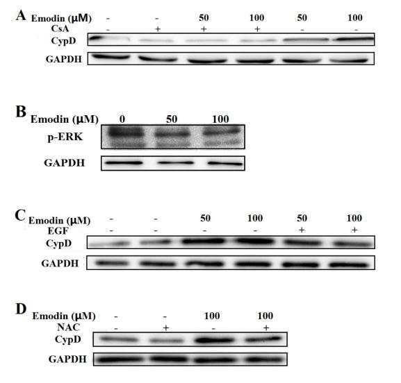
Application: WB Species: human Sample:

Application: WB Species: human Sample: HK‑2 cells
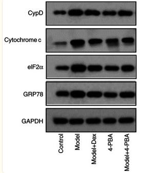
Application: WB Species: Human Sample: HK-2 cells

Application: WB Species: Human Sample: Breast Cancer MCF-7 Cells
Restrictive clause
Affinity Biosciences tests all products strictly. Citations are provided as a resource for additional applications that have not been validated by Affinity Biosciences. Please choose the appropriate format for each application and consult Materials and Methods sections for additional details about the use of any product in these publications.
For Research Use Only.
Not for use in diagnostic or therapeutic procedures. Not for resale. Not for distribution without written consent. Affinity Biosciences will not be held responsible for patent infringement or other violations that may occur with the use of our products. Affinity Biosciences, Affinity Biosciences Logo and all other trademarks are the property of Affinity Biosciences LTD.