


, diluted 1/600 was used as secondary antibody.")
 by IF/ICC. The samples were fixed with PFA and permeabilized in 0.1% Triton X-100,then blocked in 10% serum for 45 minutes at 25°C. Samples were then incubated with primary Ab(AF4039) and mouse anti-beta tubulin Ab(T0023) for 1 hour at 37°C. An AlexaFluor594 conjugated goat anti-rabbit IgG(H+L) Ab(Red) and an AlexaFluor488 conjugated goat anti-mouse IgG(H+L) Ab(Green) were used as the secondary antibody.
The nuclear counter stain is DAPI(blue).")
 Representative photographs of treated and untreated cells. PTL treatment reduced p-ERK 2, NF-κB, and Snail staining compared with sections obtained from control mice. (B and D) Representative photographs of treated and untreated cells. PTL treatment increased E-cadherin and Occludin staining and reduced Vimentin and N-cadherin staining compared with sections obtained from control mice. Each experiment was performed in triplicate. Results show the means of the three experiments, and the error bars represent standard deviation (*P < 0.05 and **P < 0.01).")
 Representative photographs of treated and untreated cells. PTL treatment reduced p-ERK 2, NF-κB, and Snail staining compared with sections obtained from control mice. (B and D) Representative photographs of treated and untreated cells. PTL treatment increased E-cadherin and Occludin staining and reduced Vimentin and N-cadherin staining compared with sections obtained from control mice. Each experiment was performed in triplicate. Results show the means of the three experiments, and the error bars represent standard deviation (*P < 0.05 and **P < 0.01).")
 The levels of cyclin A, cyclin B, cyclin D1, and CDK2 were reduced in A375 cells transfected with OVOS2-shRNA; (b) The downregulated expression of N-cadherin accompanied with the upregulated expression of E-cadherin and β-catenin were observed in A375 cells transfected with OVOS2-shRNA; (c) The expression of p-FAK, p-AKT, and p-ERK were reduced in A375 cells transfected with OVOS2-shRNA; (d) The increased production of MMP-2 was observed in A375 transfected with OVOS2-shRNA; (e) GAPDH was used as the reference.")
 and Western blot (B and C) indicated that transient
transfection of HTR8/SVneo cells with miR-210 mimic increases E-cadherin expression and decreases N-cadherin and vimentin expression at the transcriptional and
protein levels compared to those in the cells transfected with miR-210 mimic NC. qRT-PCR (E and H) and Western blot (F and G) indicated that silencing of miR-210
in HTR8/SVneo cells got the opposite results. β-actin served as the loading control. (*P < 0.05, **P < 0.01, ***P < 0.001). n = 3.")
 Overexpression of KRT7 increased Snail, vimentin and N-cadherin expression, and decreased E-cadherin expression in HEY cells as detected by western blotting. (C and D) Knockdown of KRT7 resulted in increased E-cadherin expression and reduced vimentin and Snail expression in OVCAR433 cells as detected by western blotting. All experiments were performed at least three times. Results are presented as the mean ± standard deviation. **P<0.01. KRT7, keratin 7; sh, short hairpin RNA; NC, negative control.")
 DDTC increased the protein expression level of E-cadherin and decreased the protein expression level of N-cadherin and Vimentin in SKOV3 cells.")
 The downregulation of NEAT1 in si-NEAT1 cell lines (A549 and H460) detected by qRT-PCR. (B, C) The
cell proliferation of A549 and H460 cells with si-NEAT1 measured by CCK8 assay. (D) The regulation of si-NEAT1 on
cell apoptosis tested by flow cytometry. (E, F) Transwell experiments on cell migration, invasion of si-NEAT1 cell lines
(A549 and H460). (G) The expression of EMT marker protein by Western blot normalized to GAPDH. *p < 0.05. CCK8,
Cell Counting Kit-8; EMT, epithelial–mesenchymal transition; qRT-PCR, quantitative real-time polymerase chain
reaction.")
 Relative LINP1 expression in EC9706 sh1, sh2, sh3, and sh4 was verified by qRT-PCR. The LINP1 sh2 cell line had the most significant knockdown effect compared with the control cell line (61.75%, P<0.001). (B) The Alamar Blue proliferation assay indicated that the proliferation of shRNA-LINP1 cells was significantly inhibited compared with that of the corresponding control cells and shRNA-scr cells (P<0.001). (C,D) Colony formation assays showing that the number of colonies formed was significantly lower in shRNA-LINP1 cells than in NC and shRNA-scr cells in macroscopic view (71.7 vs. 73.3 vs. 24.7, P<0.001). (E,F) Wound healing assays showed that the migration rate was slower in shRNA-LINP1 cells than in NC and shRNA-scr cells after 48 h (19.1% vs. 46.4% vs. 46.6%; P<0.001) (magnification 100×). (G,H) Transwell migration assays demonstrated that the number of migratory cells was lower in shRNA-LINP1 than in the two control cells (60.3 vs. 236.3 vs. 238.7; P<0.001) (magnification 100×). (I,J,K) qRT-PCR and western blot analyses of the expression of EMT markers. E-cadherin was significantly upregulated, whereas N-cadherin, vimentin, snail and slug were significantly downregulated in shRNA-LINP1 cells compared with NC and shRNA-scr cells (all P<0.05). **, P<0.01; ***, P<0.001.")
 qRT-PCR showing the knock down of lncRNA AL161431.1, and the corresponding changes in E-cadherin, N-cadherin, and vimentin in SW1990 and BxPC-3 cells; (B) Western blots showing the changes in protein level in E-cadherin, N-cadherin, and vimentin in SW1990 and BxPC-3 cells after transfection of scrambled siRNA or siRNA1; (C) Immunofluorescence analysis showing the change of expression of E-cadherin, N-cadherin, and vimentin in SW1990 and BxPC-3 cells after transfection of scrambled siRNA or siRNA1 (200x). *p < 0.05, **p < 0.01, ***p < 0.001.")
 qRT-PCR showing the knock down of lncRNA AL161431.1, and the corresponding changes in E-cadherin, N-cadherin, and vimentin in SW1990 and BxPC-3 cells; (B) Western blots showing the changes in protein level in E-cadherin, N-cadherin, and vimentin in SW1990 and BxPC-3 cells after transfection of scrambled siRNA or siRNA1; (C) Immunofluorescence analysis showing the change of expression of E-cadherin, N-cadherin, and vimentin in SW1990 and BxPC-3 cells after transfection of scrambled siRNA or siRNA1 (200x). *p < 0.05, **p < 0.01, ***p < 0.001.")
 NSE overexpression promoted the EMT process of SCLC. (A) Western blot assay was performed to measure the protein expression levels of the EMT-related markers (Snail, N-cadherin and E-cadherin).")
immunofluorescence analysis showing the change of expression of E-cadherin, N-cadherin, and vimentin in SW1990 and BxPC-3 cells after transfection of scrambled siRNA or siRNA1 (200×). *P<0.05, **P<0.01.")
 western blots showing the changes in protein level in E-cadherin, N-cadherin, and vimentin in SW1990 and BxPC-3 cells after transfection of scrambled siRNA or siRNA1")
 NUDCD1 knockdown decreased the expression of Ncadherin and vimentin and upregulated the expression of E-cadherin in PANC-1 cells.")
 NSE overexpression promoted the EMT process of SCLC. (A) Western blot assay was performed to measure the protein expression levels of the EMT-related markers (Snail, N-cadherin and E-cadherin). (B) qRT-PCR was performed to measure the mRNA expression levels of the EMT markers. (C, D) NSE knockdown represses the EMT process of SCLC. Protein levels (C) and mRNA levels (D) of EMT-related markers were measured using western blot or qRT-PCR, respectively. These results were repeated of three independent experiments.")
 qRT-PCR showing the knock down of lncRNA ELFN1-AS1, and the corresponding changes in E-cadherin, N-cadherin, and vimentin in SW1990 and BxPC-3 cells; (B) western blots showing the changes in protein level in E-cadherin, N-cadherin, and vimentin in SW1990 and BxPC-3 cells after transfection of scrambled siRNA or siRNA1; (C) immunofluorescence analysis showing the change of expression of E-cadherin, N-cadherin, and vimentin in SW1990 and BxPC-3 cells after transfection of scrambled siRNA or siRNA1 (200×). *P<0.05, **P<0.01.")
 NUDCD1 knockdown decreased the expression of N-cadherin and vimentin and upregulated the expression of E-cadherin in PANC-1 cells. (E–H) NUDCD1 knockdown decreased the expression of N-cadherin and vimentin, and upregulated the expression of E-cadherin in Patu8988 cells. ***p<0.001, ****p<0.0001.")
. (B) The relative protein express levels of NSE were measured by western blot. And quantification of the relative protein amount was shown in the lower panel. (C) Real time quantitative PCR confirmed the expression of NSE after being overexpressed or knockdown. (D) The effect of NSE on the proliferation capabilities of SCLC cells were assessed using CCK8 assay. Transwell assay was performed to investigate the effect of NSE on the migration and invasion of SCLC cells. (E) Overexpression of NSE promoted cell migration and invasion of H446 cells. (F) Knockdown of NSE significantly inhibited cell migration. (G) Western blot results demonstrated overexpression of NSE promoted the EMT process by down-regulating the expression of E-cadherin and upregulating the expression of N-cadherin, while silencing NSE upregulated the expression of E-cadherin and downregulated the expression of N-cadherin. And quantification of the relative protein amount was shown in the lower panel.")
 Cell migratory abilities were assessed via the wound-healing assay (scale bar = 200 μm). (b, c) Migratory and invasive abilities was examined using Transwell assay (scale bar = 50 μm). (d) Protein levels were estimated by Western blot. ∗A significant difference compared with the sh-NC group. ∗∗P < 0.01.")
 Overexpression of LAMC2 promoted the expression of N-cadherin and vimentin proteins in TU177 cells and inhibited the expression of E-cadherin protein. Western blot was performed to detect the effect of LAMC2 overexpression on the EMT in LSCC cells. (b) LAMC2 knockdown inhibited N-cadherin and vimentin protein expression and promoted E-cadherin protein expression in AMC-HN-8 cells. Western blot was performed to detect the effect of LAMC2 knockdown on the EMT in LSCC cells. Data were expressed as mean ± SD, n = 3. Compared to the vector group, ##P < 0.01. Compared to the shNC group, ∗∗P < 0.01.")
 and (b). The BHLHE41, hypoxia-inducible factor-1alpha (HIF-1α), and epithelial-mesenchymal transition- (EMT-) related factor levels in hypoxia-induced CC cells were tested by Western blot. All experiments have been performed in triplicate, and data were expressed as mean ± SD. ∗∗P < 0.01 vs. hypoxia;")
 and those cultured in regular media for the same course (Ctrl) were analyzed. (A) CCK-8 assay was applied to assess cell viability for 6 days. (B) The migration of cells was studied and quantified by transwell assay. (C) Cytotoxicity assay was used to examine apoptosis after 3 days of growth factor deprivation stress (Q3). (D) Western blot analysis showed altered expression of apoptotic markers in cells subjected to 5 days of growth factor deprivation (Q5). (E) Flow cytometry analysis was performed to study cell death in response to 48 h treatment of 5-FU (6 μM). (F) Protein levels of E-cadherin, N-cadherin, and vimentin were determined by Western blot analysis. Data were representatives of at least three independent experiments, shown as mean ± SD. Significance threshold determined by one-way ANOVA or Student's t-test:")
, collagen I (b), vimentin (c), N-cadherin (d), and a-SMA (e) protein and their protein band (f) in the lung tissue of mice in each group. NC, normal control group; BLM, bleomycin-induced systemic sclerosis model group; PESV-L, low-dose PESV intervention group; PESV-M, medium-dose PESV intervention group; PESV-H, high-dose PESV intervention group; DXM, dexamethasone intervention group.")
 NSE overexpression promoted the EMT process of SCLC. (A) Western blot assay was performed to measure the protein expression levels of the EMT-related markers (Snail, N-cadherin and E-cadherin). (B) qRT-PCR was performed to measure the mRNA expression levels of the EMT markers. (C, D) NSE knockdown represses the EMT process of SCLC. Protein levels (C) and mRNA levels (D) of EMT-related markers were measured using western blot or qRT-PCR, respectively. These results were repeated of three independent experiments.")
, scale bar = 200 μm; cell invasion using Transwell pre-coated with Matrigel (c-d), scale bar = 100 μm; the protein levels of N-cadherin, MMP2, MMP9, Vimentin, and E-cadherin using Immunoblotting (e-f). All N = 3; * P")
 Tspan5 triggers the rearrangement of actin cytoskeleton. Stress fibres and actin filaments were visualized by Rhodamine-phalloidin staining (red) in HCC cells transduced with lentivirus containing Tspan5/shTspan5-encoding vector or empty/scramble RNA vector (Control/shControl). Nuclei were stained with DAPI (blue). Upregulation of Tspan5 significantly increased F-actin expression (red) and actin stress fibres throughout the Tspan5-upregulating cells compared with the control cells in which the actin bundles were predominantly localized underneath cell membranes. 400× magnification, scale bar: 10 μm. Mean ± SD, n = 3, Student's t-test; *P < 0.05, **P < 0.01. (B) Western blotting showing regulation of the expression of EMT markers, E-cadherin, N-cadherin, vimentin and Snail by Tspan5. GAPDH was used as a protein loading control. Numbers indicating relative protein ratio measured by Image J software and normalized to GAPDH. Vimentin was undetectable in MHCC97L cells. (C) Representative IF images showing regulation of the expressions of E-cadherin and vimentin by Tspan5. 630× magnifications, scale bar: 10 μm. Quantification of the protein expression was performed by Zeiss zen microscope software. Mean ± SD, n = 9, Student's t-test; **P < 0.01. (D) Representative IHC images of Tspan5, E-cadherin and vimentin expressions in xenograft sections of metastatic foci in mouse lungs. 600× magnifications, scale bar: 50 μm. Mean ± SD,")
 Tspan5 triggers the rearrangement of actin cytoskeleton. Stress fibres and actin filaments were visualized by Rhodamine-phalloidin staining (red) in HCC cells transduced with lentivirus containing Tspan5/shTspan5-encoding vector or empty/scramble RNA vector (Control/shControl). Nuclei were stained with DAPI (blue). Upregulation of Tspan5 significantly increased F-actin expression (red) and actin stress fibres throughout the Tspan5-upregulating cells compared with the control cells in which the actin bundles were predominantly localized underneath cell membranes. 400× magnification, scale bar: 10 μm. Mean ± SD, n = 3, Student's t-test; *P < 0.05, **P < 0.01. (B) Western blotting showing regulation of the expression of EMT markers, E-cadherin, N-cadherin, vimentin and Snail by Tspan5. GAPDH was used as a protein loading control. Numbers indicating relative protein ratio measured by Image J software and normalized to GAPDH. Vimentin was undetectable in MHCC97L cells. (C) Representative IF images showing regulation of the expressions of E-cadherin and vimentin by Tspan5. 630× magnifications, scale bar: 10 μm. Quantification of the protein expression was performed by Zeiss zen microscope software. Mean ± SD, n = 9, Student's t-test; **P < 0.01. (D) Representative IHC images of Tspan5, E-cadherin and vimentin expressions in xenograft sections of metastatic foci in mouse lungs. 600× magnifications, scale bar: 50 μm. Mean ± SD,")

, the levels of E-cadherin and vimentin by immunofluorescence staining (B,C), the protein levels of E-cadherin, N-cadherin, vimentin, and snail by western blot analysis (D), as well as the protein levels of Wnt1, c-Myc, and cyclin D1 by western blot analysis (E). **P")

/targeting protein for Xklp2 (TPX2) or co-knockdown in SW480; B: AURKA/TPX2 knockdown or co-knockdown LOVO cells decreased N-cadherin, vimentin, AURKA, phosphorylated AURKA, and TPX2 expression, and increased E-cadherin expression. Epithelial-mesenchymal transition biomarkers include N-cadherin, E-cadherin, and vimentin. Western blotting was used to detect relative protein expression. 1P < 0.05 vs negative control of short hairpin RNA (sh-NC) group. 2P < 0.01 vs sh-NC group. 3P < 0.05 vs short hairpin RNA of AURKA (sh-AURKA) group. 4P < 0.01 vs sh-AURKA group. 5P < 0.05 vs short hairpin-TPX2 (sh-TPX2) group. 6P < 0.01 vs sh-TPX2 group. AURKA: Aurora kinase A; TPX2: Targeting protein for Xklp2; sh-NC: Negative control of short hairpin RNA; sh-AURKA: Short hairpin RNA of Aurora kinase A; sh-TPX2: Short hairpin RNA of targeting protein for Xklp2; p-AURKA: Phosphorylated Aurora kinase A.")
 Gene set enrichment analysis based on TCGA, GSE16011 datasets. (C–E) Protein expression of mesenchymal transformation related proteins (N-Cadherin, E-Cadherin) and PI3K/AKT/mTORC1 pathway in TJ905 and A172 cells with ITGA5 knockdown was detected by Western blot.")
| 製品: | N Cadherin Antibody |
| カタログ: | AF4039 |
| タンパク質の説明: | Rabbit polyclonal antibody to N Cadherin |
| アプリケーション: | WB IHC IF/ICC |
| Cited expt.: | WB, IHC, IF/ICC |
| 反応性: | Human, Mouse, Rat |
| 予測: | Pig, Zebrafish, Bovine, Horse, Sheep, Rabbit, Dog, Chicken, Xenopus |
| 分子量: | 140kd(Observed); 100kD(Calculated). |
| ユニプロット: | P19022 |
| RRID: | AB_2835344 |
Control Products

製品説明
*The optimal dilutions should be determined by the end user. For optimal experimental results, antibody reuse is not recommended.
*Tips:
WB: For western blot detection of denatured protein samples. IHC: For immunohistochemical detection of paraffin sections (IHC-p) or frozen sections (IHC-f) of tissue samples. IF/ICC: For immunofluorescence detection of cell samples. ELISA(peptide): For ELISA detection of antigenic peptide.
引用形式: Affinity Biosciences Cat# AF4039, RRID:AB_2835344.
折りたたみ/展開
CADH2_HUMAN; Cadherin 2; Cadherin 2 N cadherin neuronal; Cadherin 2 type 1; Cadherin 2 type 1 N cadherin neuronal; Cadherin 2, type 1, N-cadherin (neuronal); Cadherin-2; Cadherin2; Calcium dependent adhesion protein neuronal; CD325; CD325 antigen; CDH2; CDHN; CDw325; CDw325 antigen; N cadherin 1; N-cadherin; NCAD; Neural cadherin; OTTHUMP00000066304; OTTHUMP00000067378;
免疫原
A synthesized peptide derived from human N Cadherin, corresponding to a region within C-terminal amino acids.
- P19022 CADH2_HUMAN:
- Protein BLAST With
- NCBI/
- ExPASy/
- Uniprot
MCRIAGALRTLLPLLAALLQASVEASGEIALCKTGFPEDVYSAVLSKDVHEGQPLLNVKFSNCNGKRKVQYESSEPADFKVDEDGMVYAVRSFPLSSEHAKFLIYAQDKETQEKWQVAVKLSLKPTLTEESVKESAEVEEIVFPRQFSKHSGHLQRQKRDWVIPPINLPENSRGPFPQELVRIRSDRDKNLSLRYSVTGPGADQPPTGIFIINPISGQLSVTKPLDREQIARFHLRAHAVDINGNQVENPIDIVINVIDMNDNRPEFLHQVWNGTVPEGSKPGTYVMTVTAIDADDPNALNGMLRYRIVSQAPSTPSPNMFTINNETGDIITVAAGLDREKVQQYTLIIQATDMEGNPTYGLSNTATAVITVTDVNDNPPEFTAMTFYGEVPENRVDIIVANLTVTDKDQPHTPAWNAVYRISGGDPTGRFAIQTDPNSNDGLVTVVKPIDFETNRMFVLTVAAENQVPLAKGIQHPPQSTATVSVTVIDVNENPYFAPNPKIIRQEEGLHAGTMLTTFTAQDPDRYMQQNIRYTKLSDPANWLKIDPVNGQITTIAVLDRESPNVKNNIYNATFLASDNGIPPMSGTGTLQIYLLDINDNAPQVLPQEAETCETPDPNSINITALDYDIDPNAGPFAFDLPLSPVTIKRNWTITRLNGDFAQLNLKIKFLEAGIYEVPIIITDSGNPPKSNISILRVKVCQCDSNGDCTDVDRIVGAGLGTGAIIAILLCIIILLILVLMFVVWMKRRDKERQAKQLLIDPEDDVRDNILKYDEEGGGEEDQDYDLSQLQQPDTVEPDAIKPVGIRRMDERPIHAEPQYPVRSAAPHPGDIGDFINEGLKAADNDPTAPPYDSLLVFDYEGSGSTAGSLSSLNSSSSGGEQDYDYLNDWGPRFKKLADMYGGGDD
種類予測
Score>80(red) has high confidence and is suggested to be used for WB detection. *The prediction model is mainly based on the alignment of immunogen sequences, the results are for reference only, not as the basis of quality assurance.
High(score>80) Medium(80>score>50) Low(score<50) No confidence
研究背景
Calcium-dependent cell adhesion protein; preferentially mediates homotypic cell-cell adhesion by dimerization with a CDH2 chain from another cell. Cadherins may thus contribute to the sorting of heterogeneous cell types. Acts as a regulator of neural stem cells quiescence by mediating anchorage of neural stem cells to ependymocytes in the adult subependymal zone: upon cleavage by MMP24, CDH2-mediated anchorage is affected, leading to modulate neural stem cell quiescence. CDH2 may be involved in neuronal recognition mechanism. In hippocampal neurons, may regulate dendritic spine density.
Cleaved by MMP24. Ectodomain cleavage leads to the generation of a soluble 90 kDa amino-terminal soluble fragment and a 45 kDa membrane-bound carboxy-terminal fragment 1 (CTF1), which is further cleaved by gamma-secretase into a 35 kDa. Cleavage in neural stem cells by MMP24 affects CDH2-mediated anchorage of neural stem cells to ependymocytes in the adult subependymal zone, leading to modulate neural stem cell quiescence (By similarity).
May be phosphorylated by OBSCN.
Cell membrane>Single-pass type I membrane protein. Cell membrane>Sarcolemma. Cell junction. Cell surface.
Note: Colocalizes with TMEM65 at the intercalated disk in cardiomyocytes. Colocalizes with OBSCN at the intercalated disk and at sarcolemma in cardiomyocytes.
Three calcium ions are usually bound at the interface of each cadherin domain and rigidify the connections, imparting a strong curvature to the full-length ectodomain. Calcium-binding sites are occupied sequentially in the order of site 3, then site 2 and site 1.
研究領域
· Environmental Information Processing > Signaling molecules and interaction > Cell adhesion molecules (CAMs). (View pathway)
· Human Diseases > Cardiovascular diseases > Arrhythmogenic right ventricular cardiomyopathy (ARVC).
参考文献

Application: WB Species: human Sample: BPH-1 cells

Application: WB Species: human Sample: breast cancer cells

Application: WB Species: Human Sample: breast cancer cells
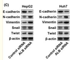
Application: WB Species: Human Sample: HepG2 and Huh7 cells

Application: WB Species: human Sample: L02, HepG2, Huh7, and MHCC97H cells

Application: WB Species: Sample: RSCs

Application: IF/ICC Species: Sample: RSCs
Restrictive clause
Affinity Biosciences tests all products strictly. Citations are provided as a resource for additional applications that have not been validated by Affinity Biosciences. Please choose the appropriate format for each application and consult Materials and Methods sections for additional details about the use of any product in these publications.
For Research Use Only.
Not for use in diagnostic or therapeutic procedures. Not for resale. Not for distribution without written consent. Affinity Biosciences will not be held responsible for patent infringement or other violations that may occur with the use of our products. Affinity Biosciences, Affinity Biosciences Logo and all other trademarks are the property of Affinity Biosciences LTD.