製品: Vimentin Antibody
カタログ: AF0292
タンパク質の説明: Rabbit polyclonal antibody to Vimentin
アプリケーション: WB IHC IF/ICC
Cited expt.: WB, IHC
反応性: Human, Mouse, Rat
予測: Pig, Bovine, Sheep, Rabbit, Dog, Chicken
分子量: 53kDa; 54kD(Calculated).
ユニプロット: P08670
RRID: AB_2833458

類似製品を見る>>

   サイズ 価格 在庫状況
 100ul $280 在庫あり
 200ul $350 在庫あり

リードタイム: 当日配達

For pricing and ordering contact:
お問い合わせ先

製品説明

ソース:
Rabbit
アプリケーション:
WB 1:500-1:3000, IHC 1:50-1:200, IF/ICC 1:100-1:500
*The optimal dilutions should be determined by the end user. For optimal experimental results, antibody reuse is not recommended.
*Tips:

WB: For western blot detection of denatured protein samples. IHC: For immunohistochemical detection of paraffin sections (IHC-p) or frozen sections (IHC-f) of tissue samples. IF/ICC: For immunofluorescence detection of cell samples. ELISA(peptide): For ELISA detection of antigenic peptide.

反応性:
Human,Mouse,Rat
予測:
Pig(100%), Bovine(100%), Sheep(100%), Rabbit(100%), Dog(100%), Chicken(100%)
クローナリティ:
Polyclonal
特異性:
Vimentin Antibody detects endogenous levels of total Vimentin.
RRID:
AB_2833458
引用形式: Affinity Biosciences Cat# AF0292, RRID:AB_2833458.
コンジュゲート:
Unconjugated.
精製:
The antiserum was purified by peptide affinity chromatography using SulfoLink™ Coupling Resin (Thermo Fisher Scientific).
保存:
Rabbit IgG in phosphate buffered saline , pH 7.4, 150mM NaCl, 0.02% sodium azide and 50% glycerol. Store at -20 °C. Stable for 12 months from date of receipt.
別名:

折りたたみ/展開

CTRCT30; Epididymis luminal protein 113; FLJ36605; HEL113; VIM; VIME_HUMAN; Vimentin;

免疫原

免疫原:

A synthesized peptide derived from human Vimentin, corresponding to a region within N-terminal amino acids.

Uniprot:
遺伝子(ID):
発現特異性:
P08670 VIME_HUMAN:

Highly expressed in fibroblasts, some expression in T- and B-lymphocytes, and little or no expression in Burkitt's lymphoma cell lines. Expressed in many hormone-independent mammary carcinoma cell lines.

タンパク質の説明:
vimentin an intermediate filament protein. Intermediate filament proteins are expressed in a tissue-specific manner. Desmin is the subunit specific for muscle and vimentin the subunit specific for mesenchymal tissue.
タンパク質配列:
MSTRSVSSSSYRRMFGGPGTASRPSSSRSYVTTSTRTYSLGSALRPSTSRSLYASSPGGVYATRSSAVRLRSSVPGVRLLQDSVDFSLADAINTEFKNTRTNEKVELQELNDRFANYIDKVRFLEQQNKILLAELEQLKGQGKSRLGDLYEEEMRELRRQVDQLTNDKARVEVERDNLAEDIMRLREKLQEEMLQREEAENTLQSFRQDVDNASLARLDLERKVESLQEEIAFLKKLHEEEIQELQAQIQEQHVQIDVDVSKPDLTAALRDVRQQYESVAAKNLQEAEEWYKSKFADLSEAANRNNDALRQAKQESTEYRRQVQSLTCEVDALKGTNESLERQMREMEENFAVEAANYQDTIGRLQDEIQNMKEEMARHLREYQDLLNVKMALDIEIATYRKLLEGEESRISLPLPNFSSLNLRETNLDSLPLVDTHSKRTLLIKTVETRDGQVINETSQHHDDLE

種類予測

種類予測:

Score>80(red) has high confidence and is suggested to be used for WB detection. *The prediction model is mainly based on the alignment of immunogen sequences, the results are for reference only, not as the basis of quality assurance.

Species
Results
Score
Pig
100
Bovine
100
Sheep
100
Dog
100
Chicken
100
Rabbit
100
Horse
0
Xenopus
0
Zebrafish
0
Model Confidence:
High(score>80) Medium(80>score>50) Low(score<50) No confidence

研究背景

機能:

Vimentins are class-III intermediate filaments found in various non-epithelial cells, especially mesenchymal cells. Vimentin is attached to the nucleus, endoplasmic reticulum, and mitochondria, either laterally or terminally.

Involved with LARP6 in the stabilization of type I collagen mRNAs for CO1A1 and CO1A2.

PTMs:

Filament disassembly during mitosis is promoted by phosphorylation at Ser-55 as well as by nestin (By similarity). One of the most prominent phosphoproteins in various cells of mesenchymal origin. Phosphorylation is enhanced during cell division, at which time vimentin filaments are significantly reorganized. Phosphorylation by PKN1 inhibits the formation of filaments. Phosphorylated at Ser-56 by CDK5 during neutrophil secretion in the cytoplasm. Phosphorylated by STK33. Phosphorylated on tyrosine residues by SRMS.

O-glycosylated during cytokinesis at sites identical or close to phosphorylation sites, this interferes with the phosphorylation status.

S-nitrosylation is induced by interferon-gamma and oxidatively-modified low-densitity lipoprotein (LDL(ox)) possibly implicating the iNOS-S100A8/9 transnitrosylase complex.

細胞の位置付け:

Cytoplasm. Cytoplasm>Cytoskeleton. Nucleus matrix.

Extracellular region or secreted Cytosol Plasma membrane Cytoskeleton Lysosome Endosome Peroxisome ER Golgi apparatus Nucleus Mitochondrion Manual annotation Automatic computational assertionSubcellular location
組織特異性:

Highly expressed in fibroblasts, some expression in T- and B-lymphocytes, and little or no expression in Burkitt's lymphoma cell lines. Expressed in many hormone-independent mammary carcinoma cell lines.

タンパク質ファミリー:

The central alpha-helical coiled-coil IF rod domain mediates elementary homodimerization.

The [IL]-x-C-x-x-[DE] motif is a proposed target motif for cysteine S-nitrosylation mediated by the iNOS-S100A8/A9 transnitrosylase complex.

Belongs to the intermediate filament family.

研究領域

· Human Diseases > Infectious diseases: Viral > Epstein-Barr virus infection.

· Human Diseases > Cancers: Overview > MicroRNAs in cancer.

参考文献

1). Stearoyl-CoA desaturase-1 promotes colorectal cancer metastasis in response to glucose by suppressing PTEN. JOURNAL OF EXPERIMENTAL & CLINICAL CANCER RESEARCH, 2018 (PubMed: 29530061) [IF=11.3]

Application: WB    Species: human    Sample: CRC

Fig. 2| SCD1 promotes migration and invasion of colorectal cancer cells by regulating EMT. i, j Protein levels of E-cadherin and vimentin in colorectal cancer cells transfected with SCD1 shRNA (i) or SCD1 cDNA (j)

2). Molecular mechanism of albumin in suppressing invasion and metastasis of hepatocellular carcinoma. LIVER INTERNATIONAL, 2022 (PubMed: 34854209) [IF=6.0]

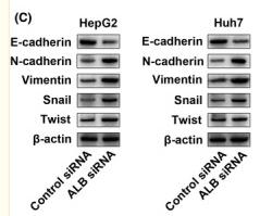
Application: WB    Species: Human    Sample: HepG2 and Huh7 cells

FIGURE 7 A, Representative images of the western blot results for uPAR, MMP2 and MMP9 in ALB knockdown HepG2 and Huh7 cells; B, Zymography analysis illustrates MMP2 and MMP9 activity in ALB knockdown HepG2 and Huh7 cells; C, Quantitative analysis results and representative images of the western blot results for the EMT‐associated markers, E‐cadherin, N‐cadherin, vimentin, Snail and Twist by western blot in ALB knockdown HepG2 and Huh7 cells; D, Quantification shows a significantly higher uPAR in HCC group with ALB <3.5 g/dL compared to ALB ≥3.5 g/dL (*P < .05); E, Scatterplot showing the correlation between plasma levels of ALB and uPAR. The vertical position represents the expression levels of uPAR (lg pg/mL)

3). Knockdown of Nucleostemin in an ovarian cancer SKOV-3 cell line and its effects on cell malignancy. BIOCHEMICAL AND BIOPHYSICAL RESEARCH COMMUNICATIONS, 2017 (PubMed: 28412352) [IF=2.5]

Application: WB    Species: human    Sample:

Fig. 3. Knockdown of NS inhibited tumor migration and invasion in vitro. (A) Crystal violet staining of the shNS and control group cells that crossed the polycarbonate membrane of the Transwell chamber to detect cell migration. (B) The number of cells that crossed the Transwell migration chamber in different groups. (C) Crystal violet staining of the shNS and control group cells that crossed the Matrigel-coated polycarbonate membrane of the Transwell chamber to detect cell invasion. (D) The number of cells that crossed the Transwell invasion chamber in different groups. (E) Representative Western blotting results indicate the EMT marker expressions in the different groups. The results are presented as the means ± SD, as based on three independent experiments. Statistical significance was determined using Student's t-test. *P < 0.05. Scale: 100 mm.

Application: IHC    Species: human    Sample:

Fig. 4. Knockdown of NS suppressed tumor formation and growth in vivo. (A) The shNS and control group cells were inoculated s.c. into the right flanks of BALB/c nude mice. Tumor volumes were monitored and recorded weekly (n ¼ 3). The results are presented as the means ± SD, as based on three independent experiments. ****P < 0.0001. (B) Examples of the tumors excised from the shNS and control mice on the 19th week following injection. Scale: 2 cm. (C) Immunohistochemistry staining of the tumor sections obtained from mice in the shNS and control groups. Antibodies: NS, Ki-67, cyclin D1, and Vimentin.

4). High glucose induces epithelial-mesenchymal transition and results in the migration and invasion of colorectal cancer cells. Experimental and Therapeutic Medicine, 2018 (PubMed: 29896243) [IF=2.4]

5). High CCL7 expression is associated with migration, invasion and bone metastasis of non-small cell lung cancer cells. American Journal of Translational Research, 2019 (PubMed: 30788000) [IF=1.7]

Application: WB    Species: mouse    Sample: A549 and PC9 cells

Figure 4. |CCL7 induces EMT in A549 and PC9 cells via interacting with CCR3. A. Protein expression of CCR1, CCR2 and CCR3 in A549 and PC9 cells with different doses of recombinant CCL7 (100 ng/ml, 200 ng/ml); the result showed that CCL7 had no effect on the expression of CCR. B-D. Wound healing assay and transwell invasion assay of A549 and PC9 cells after co-incubation with recombinant CCL7 and CCR inhibitors respectively; interestingly, inhibition of CCR3 significantly suppressed cell invasion. E. CCL7 increased the expression of mesenchymal markers and decreased the expression of epithelial markers in A549 and PC9 cells via CCR3.

6). Doxycycline directly targets PAR1 to suppress tumor progression. Oncotarget, 2017 (PubMed: 28187433)

Application: IHC    Species: human    Sample:

(F) Immunohistochemical staining to identify EMT biomarkers and doxycycline inhibited proteins in treated and untreated cells. Doxycycline-treated cells display stronger E-cadherin and α-SMA staining but reduced vimentin,MMP-2 and MMP-9 staining;

Restrictive clause


Affinity Biosciences tests all products strictly. Citations are provided as a resource for additional applications that have not been validated by Affinity Biosciences. Please choose the appropriate format for each application and consult Materials and Methods sections for additional details about the use of any product in these publications.

For Research Use Only.
Not for use in diagnostic or therapeutic procedures. Not for resale. Not for distribution without written consent. Affinity Biosciences will not be held responsible for patent infringement or other violations that may occur with the use of our products. Affinity Biosciences, Affinity Biosciences Logo and all other trademarks are the property of Affinity Biosciences LTD.