

, using Smad3 Antibody. The lane on the left was treated with blocking peptide.")
.
Bands result from membrane strip incubation.")


 and mouse anti-beta tubulin Ab(T0023 1:200) for 1 hour at 37°C. An AlexaFluor594 conjugated goat anti-rabbit IgG(H+L) Ab(Red) and an AlexaFluor488 conjugated goat anti-mouse IgG(H+L) Ab(Green) were used as the secondary antibody.
The nuclear counter stain is DAPI(blue).")
 Representative proteins were investigated using western blot analysis. The cells of two group were treated for twenty-four hours. In Fig. B and C, NC/0: primary cells, N30/30: thirty generations of bovine mammary epithelial cells treated by IFN-γ (10 ng/ml). RNAseq: transcriptomics results. qRT-PCR: fluorescence quantitative PCR results. The data represent the means ± SEM of 3 independent experiments. Each bar represents the mean of three independent experiments. One-way ANOVA; **P < 0.01.")
 and pCX43 (C) showed a significant decrease but TGF-β1 (D), Smad3 (E), and pSmad3 (F) showed a significant increase in the bladder detrusor after SCI. Those changes were more significant in transection than in hemisection of sacral spine cord. CX45 was not changed among three groups.")
 CFs were incubated without or with TGF-β1 (10 ng/ml) and
THSWD (15, 30 and 60 μg/ml) for 24 h, and the expression levels of collagen I, collagen III, collagen V, phospho-TGFBR1, TGFBR1, phospho-Smad2, Smad2,
phospho-Smad3 and Smad3 were tested by western blotting. (C–E) Expression levels of collagen I, collagen III and collagen V were normalized with GAPDH (n = 3).
(F–H) Expression levels of phospho-TGFBR1, phospho-Smad2, and phospho-Smad3 were normalized to that of TGFBR1, Smad2 and Smad3 proteins, respectively (n
= 3). Data were shown as mean ± SD. #P < 0.05, vs. control group. *P < 0.05, **P < 0.01, vs. model group.")
 IHC showed inhibition of TGFBR1, Smad3, collagen I, collagen III and α-SMA in THSWD-treated mouse heart tissues compared with the model group. (B–F)
Quantitative analysis for IHC staining of TGFBR1, Smad3, collagen I, collagen III and α-SMA. Data were shown as mean ± SD. **P < 0.01, vs. model group.")

 for 48 h before being exposed to H/R. D-F, Western blot analysis for the protein expression of ALK5, Smad2, Smad3, p-Smad2, and Smad3 in the indicated groups and quantitative analysis of ALK5, p-Smad2, and p-Smad3, n = 3.")
 Te expression levels of the renal fbrosis-associated genes TGF-β1, FN, Smad3, Col1a1, Col1a2, and α-SMA were quantitated in each of the groups. All groups were compared with OC. *stands for p<0.05, **stands for p<0.01, and ***stands for p<0.001. (b) Te western blot analysis of TGF-β1, Smad3, a-SMA, FN, Col1a1 and Col1a2.")
 Western blot analysis of the protein levels of p-Akt, p-Smad3, p-p38 and p-Erk1/2 expression.")
 Western blot results of TGF-β1, Smad2, p-Smad2, Smad3, p-Smad3 and Smad7 protein expression levels. n = 3 rats per group.")
 The representative image of flow-cytometry of CD206 expression and CD86 expression in THP-1 macrophages with different stimulations. (B) The quantification of CD206 expression in THP-1 macrophages with different stimulations. (C) The quantification of CD86 expression in THP-1 macrophages with different stimulations. (D) The expression of Smad3 and p-Smad3 in THP-1 macrophages with different stimulations, and related quantification (E and F). N=7. ***p<0.001.")
 were infected with adenovirus encoding shRNA targeting TFPI2 (TFPI2 shRNA) or overexpressing TFPI2 (TFPI2 OE), followed by stimulation of 5 ng/ml TGF-β2 for 48 h. A, the expression of SMAD7, TGFBR1, TGFBR2, SMAD2/3, and phospho-SMAD2/3 (p-SMAD2/3) was determined by Western blot. Semiquantitative analysis of (B) SMAD7, (C) TGFBR1, and (D) TGFBR2, as well as (E and F) the ratio of p-SMAD2/3 to SMAD2/3. G and H, immunofluorescent staining of SMAD2/3 in hRGECs. Yellow arrows indicated nuclear translocation of SMAD2/3. Data are shown as the mean ± SD (n = 3). ∗p < 0.05, ∗∗p < 0.01, ∗∗∗p < 0.001. TGF-β, transforming growth factor beta; TFP12, tissue factor pathway inhibitor 2.")
 were infected with adenovirus encoding shRNA targeting TFPI2 (TFPI2 shRNA) or overexpressing TFPI2 (TFPI2 OE), followed by stimulation of 5 ng/ml TGF-β2 for 48 h. A, the expression of SMAD7, TGFBR1, TGFBR2, SMAD2/3, and phospho-SMAD2/3 (p-SMAD2/3) was determined by Western blot. Semiquantitative analysis of (B) SMAD7, (C) TGFBR1, and (D) TGFBR2, as well as (E and F) the ratio of p-SMAD2/3 to SMAD2/3. G and H, immunofluorescent staining of SMAD2/3 in hRGECs. Yellow arrows indicated nuclear translocation of SMAD2/3. Data are shown as the mean ± SD (n = 3). ∗p < 0.05, ∗∗p < 0.01, ∗∗∗p < 0.001. TGF-β, transforming growth factor beta; TFP12, tissue factor pathway inhibitor 2.")

, TβRI (b), TβRII (c), p-Smad2 (d), p-Smad3 (e), and Smad7 (f) protein and their protein band ((g), (h)) in the lung tissue of mice in each group. NC, normal control group; BLM, bleomycin-induced systemic sclerosis model group; PESV-L, low-dose PESV intervention group; PESV-M, medium-dose PESV intervention group; PESV-H, high-dose PESV intervention group; DXM, dexamethasone intervention group.")
. As compared to the normal group, the smad3 and TGF-β1 proteins elevated in all the groups excepting the high dose of CA-treated group(P")
. As compared to the normal group, the smad3 and TGF-β1 proteins elevated in all the groups excepting the high dose of CA-treated group(P")

 Binding site prediction of miR-122-5p in SMAD3 3′-UTR. (B) Luciferase analysis showing miR-122-5p targeting SMAD3. (C) The expression of miR-122-5p in Exo-miR-122-5p and Exo-in-miR-122-5p. (D) Protein expression of SMAD3 and p-SMAD3 in DPCs treated with Exo-miR-122-5p and Exo-in-miR-122-5p.")
. All experiments were repeated three times.")
 ZH-254 on HEL cell cycle by flow cytometry. Statistical analysis of the proportion of cells in different cell cycle phases in different periods. (B,C) Regulation of ZH-254 on G1-phase-related gene and protein expression was assessed by RT-PCR and Western blotting. Quantitative analysis with Image J software (V1.8.0.112, NIH, Bethesda, MD USA). * p < 0.05, *** p < 0.001.")
 Cell viability was evaluated by the CCK-8 assay. (B) The qRT-PCR analysis of TGFβ1, COL1A2, and ACTA2 (α-SMA) mRNA levels of LF cells which were treated with the optimal concentration of the THBS1 protein. (C) Western blotting assay showing protein expression levels of TGFβ1, Smad3, COL1A2, and α-SMA of LF cells which were treated with various dosages of THBS1 for 24 h. (D) Immunohistochemical staining of p-Smad3 of the LF samples from LFH group (n = 6) and non-LFH group (n = 6), and the percentage of positive cells was quantified. Scale bar = 20 μm. (E) Protein levels of TGFβ1, Smad3, COL1A2, and α-SMA of LF cells with different treatments. (F) Immunofluorescence staining of p-Smad3 of LF cells with different treatments. Nuclei were stained with DAPI (blue). p-Smad3 was stained red. Scale bar = 20 μm. (G) Double-labeling immunohistofluorescence staining of TGFβ1 and COL1A2 of cells with different treatments. Nuclei were stained with DAPI (blue). TGFβ1 was stained green. COL1A2 was stained red. Scale bar = 20 μm. These cells were mechanically stretched for 16 h. THBS1 and LSKL stimulated cells for 24 h. These data are representative of three independent experiments. Error bars: mean ± S.D. ns, not significant. ***p < 0.001.")
, α-SMA (B), Erbb4-IR (C) and miR-29b (D) in different groups was detected by qRT-PCR (n = 6), the results were presented as mean ± SD, *P")
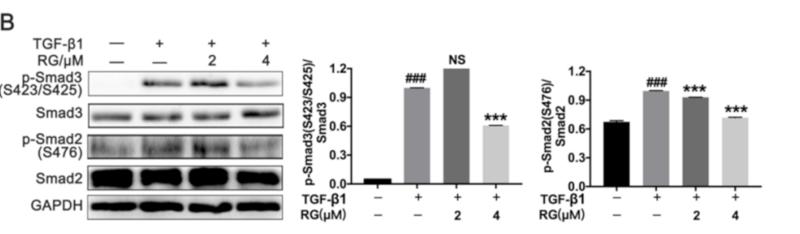
 The CAGA-NIH-3T3 cells were exposed to TGF-β1 and/or a serious concentration (0–32 µM) in serum-free medium for 18 hours. (B) The Mlg cells were treated with/without TGF-β1 and/or PZ (2 or 4 μM) for 30 minutes, and made use of Western bolt to evaluate the p-Smad3 and p-Smad2 expression levels. (C,D) Densitometric analysis of the immunoblot reported in (B). (E) Mlg cells were exposed to TGF-β1 and/or PZ (2 or 4 µM) for 12 hours, then detected for the expression of Akt, Erk and its phosphorylation by Western bolt. (F) The BLM-PPF cells were incubated with PZ (2 or 4 μM) for 24 hours. Data in (C,D) are mean ± standard deviation. ###, P")
 and MLg (B) cells were exposed to oleuropein for 12 h and then treated with TGF-β1 (5 ng/mL) for 0.5 h before samples were collected for Western blot analysis to evaluate the protein expression levels of P-Smad2, Smad2, P-Smad3, Smad3 (original images can be found in Figure S1). The data are presented as Mean ± SD (one-way ANOVA with Tukey’s post hoc multiple comparison tests), n = 3. # indicates differences between the control group and the TGF-β1 group, with # p < 0.05, ## p < 0.01, and #### p < 0.0001. * indicates differences between the TGF-β1 group and the oleuropein treatment groups, with * p < 0.05, ** p < 0.01, *** p < 0.001, and **** p < 0.0001.")
 Representative images showing mouse hearts. Bar = 50 mm. (B) Representative hematoxylin and eosin (H&E) staining images. Bar = 200 μm. (C) Representative Wheat germ agglutinin (WGA) staining images. Bar = 20 μm. (D) Quantification cross-sectional area in (G); n = 100 cells per group. (E) Representative Masson’s trichrome staining images. Bar = 50 μm. (F) Quantification percentage of interstitial fibrosis in (E). (G) and (I) Representative images of myocardial type I collagen (collagen I, Col I) and type III collagen (collagen III, Col III) immunohistochemistry (IHC). Bar = 200 μm. (H) and (J) IHC quantification of collagen I and collagen III. (K) Representative western blot images of Col I, Col III, p-Smad2, and p-Smad3 expression in the left-ventricular samples from each group of mice. (L) Quantification of the protein expression of TGFBR1 in (K). Mean ± SEM, n = 6 mice per group for panels A–L. The normality of data distribution was tested using the Shapiro–Wilk method. One-way ANOVA was applied in (D), (F), (H), (J), and (L). *p")
 and Smad3 (right) in Normal/Cancer, as analyzed using bioinformatics platforms (GEPIA and XIANTAO). *P")

 RT-qPCR detection of mRNA expression of components in TGF-β/Smad signaling and collagen genes. (B, C) Representative Western blot results (B) and quantification (C) of protein expression of components in TGF-β/Smad signaling and collagen proteins.")
 Western blot analysis of TGF-β1, TGF-βR1, p-Smad2, Smad2, p-Smad3, and Smad3 protein levels (fold change normalized to GAPDH or total Smad2/3) in lung tissues from sham, COPD, and COPD + OMT mice. (F) Serum TGF-β1 concentration (ng/mL) quantified by ELISA. N = 8 mice/group. (G) TGF-β1 secretion (ng/mL) in HBE cell supernatants after 10% CSE (v/v, 48 h) ± OMT (80 μM, 24-h pretreatment). (H–I) Time-dependent phosphorylation of Smad2/3 (fold change normalized to total Smad2/3) in HBE cells exposed to 10% CSE (v/v, 0–60 min) ± OMT (80 μM, 24-h pretreatment). (J–L) Immunofluorescence staining (scale bar: 50 μm) and quantification of nuclear Smad2/3 intensity (RFU) in HBE cells treated with 10% CSE (v/v, 30 min) ± OMT (80 μM, 24-h pretreatment). N = 3. ###p")
. B Immunofluorescence analysis of α-SMA. scale bar = 20 μm. (n = 6). C mRNA levels of fibrogenic markers (α-Sma, Col1a1, and Timp1). (n = 6) *P < 0.05, **P < 0.01 compared with si-NC + TGF-β1; $P < 0.05, $$P < 0.01 compared with si-NC; #P < 0.05, ##P < 0.01 compared with si-VDR + TGF-β1")
 Representative images of protein bands. (b-e) TGF-β1, (c) p-ERK1/2 / ERK1/2, (d) p-Smad2/Smad2, (e) pSmad3/Smad3, (f) type II collagen, (g) ALP, (h) TRACP, and (i) BMP7 were measured by western blot. (n=3, means ± SD) **P")
. β-actin as a loading control")

製品説明
*The optimal dilutions should be determined by the end user. For optimal experimental results, antibody reuse is not recommended.
*Tips:
WB: For western blot detection of denatured protein samples. IHC: For immunohistochemical detection of paraffin sections (IHC-p) or frozen sections (IHC-f) of tissue samples. IF/ICC: For immunofluorescence detection of cell samples. ELISA(peptide): For ELISA detection of antigenic peptide.
引用形式: Affinity Biosciences Cat# AF6362, RRID:AB_2835210.
折りたたみ/展開
DKFZP586N0721; DKFZp686J10186; hMAD 3; hMAD-3; hSMAD3; HSPC193; HST17436; JV15 2; JV15-2; JV152; LDS1C; LDS3; MAD (mothers against decapentaplegic Drosophila) homolog 3; MAD homolog 3; Mad homolog JV15 2; Mad protein homolog; MAD, mothers against decapentaplegic homolog 3; Mad3; MADH 3; MADH3; MGC60396; Mothers against decapentaplegic homolog 3; Mothers against DPP homolog 3; SMA and MAD related protein 3; SMAD 3; SMAD; SMAD family member 3; SMAD, mothers against DPP homolog 3; Smad3; SMAD3_HUMAN;
免疫原
A synthesized peptide derived from human Smad3, corresponding to a region within C-terminal amino acids.
- P84022 SMAD3_HUMAN:
- Protein BLAST With
- NCBI/
- ExPASy/
- Uniprot
MSSILPFTPPIVKRLLGWKKGEQNGQEEKWCEKAVKSLVKKLKKTGQLDELEKAITTQNVNTKCITIPRSLDGRLQVSHRKGLPHVIYCRLWRWPDLHSHHELRAMELCEFAFNMKKDEVCVNPYHYQRVETPVLPPVLVPRHTEIPAEFPPLDDYSHSIPENTNFPAGIEPQSNIPETPPPGYLSEDGETSDHQMNHSMDAGSPNLSPNPMSPAHNNLDLQPVTYCEPAFWCSISYYELNQRVGETFHASQPSMTVDGFTDPSNSERFCLGLLSNVNRNAAVELTRRHIGRGVRLYYIGGEVFAECLSDSAIFVQSPNCNQRYGWHPATVCKIPPGCNLKIFNNQEFAALLAQSVNQGFEAVYQLTRMCTIRMSFVKGWGAEYRRQTVTSTPCWIELHLNGPLQWLDKVLTQMGSPSIRCSSVS
種類予測
Score>80(red) has high confidence and is suggested to be used for WB detection. *The prediction model is mainly based on the alignment of immunogen sequences, the results are for reference only, not as the basis of quality assurance.
High(score>80) Medium(80>score>50) Low(score<50) No confidence
研究背景
Receptor-regulated SMAD (R-SMAD) that is an intracellular signal transducer and transcriptional modulator activated by TGF-beta (transforming growth factor) and activin type 1 receptor kinases. Binds the TRE element in the promoter region of many genes that are regulated by TGF-beta and, on formation of the SMAD3/SMAD4 complex, activates transcription. Also can form a SMAD3/SMAD4/JUN/FOS complex at the AP-1/SMAD site to regulate TGF-beta-mediated transcription. Has an inhibitory effect on wound healing probably by modulating both growth and migration of primary keratinocytes and by altering the TGF-mediated chemotaxis of monocytes. This effect on wound healing appears to be hormone-sensitive. Regulator of chondrogenesis and osteogenesis and inhibits early healing of bone fractures. Positively regulates PDPK1 kinase activity by stimulating its dissociation from the 14-3-3 protein YWHAQ which acts as a negative regulator.
Phosphorylated on serine and threonine residues. Enhanced phosphorylation in the linker region on Thr-179, Ser-204 and Ser-208 on EGF and TGF-beta treatment. Ser-208 is the main site of MAPK-mediated phosphorylation. CDK-mediated phosphorylation occurs in a cell-cycle dependent manner and inhibits both the transcriptional activity and antiproliferative functions of SMAD3. This phosphorylation is inhibited by flavopiridol. Maximum phosphorylation at the G(1)/S junction. Also phosphorylated on serine residues in the C-terminal SXS motif by TGFBR1 and ACVR1. TGFBR1-mediated phosphorylation at these C-terminal sites is required for interaction with SMAD4, nuclear location and transactivational activity, and appears to be a prerequisite for the TGF-beta mediated phosphorylation in the linker region. Dephosphorylated in the C-terminal SXS motif by PPM1A. This dephosphorylation disrupts the interaction with SMAD4, promotes nuclear export and terminates TGF-beta-mediated signaling. Phosphorylation at Ser-418 by CSNK1G2/CK1 promotes ligand-dependent ubiquitination and subsequent proteasome degradation, thus inhibiting SMAD3-mediated TGF-beta responses. Phosphorylated by PDPK1.
Acetylation in the nucleus by EP300 in the MH2 domain regulates positively its transcriptional activity and is enhanced by TGF-beta.
Poly-ADP-ribosylated by PARP1 and PARP2. ADP-ribosylation negatively regulates SMAD3 transcriptional responses during the course of TGF-beta signaling.
Ubiquitinated. Monoubiquitinated, leading to prevent DNA-binding. Deubiquitination by USP15 alleviates inhibition and promotes activation of TGF-beta target genes. Ubiquitinated by RNF111, leading to its degradation: only SMAD3 proteins that are 'in use' are targeted by RNF111, RNF111 playing a key role in activating SMAD3 and regulating its turnover (By similarity). Undergoes STUB1-mediated ubiquitination and degradation.
Cytoplasm. Nucleus.
Note: Cytoplasmic and nuclear in the absence of TGF-beta. On TGF-beta stimulation, migrates to the nucleus when complexed with SMAD4 (PubMed:15799969). Through the action of the phosphatase PPM1A, released from the SMAD2/SMAD4 complex, and exported out of the nucleus by interaction with RANBP1 (PubMed:16751101, PubMed:19289081). Co-localizes with LEMD3 at the nucleus inner membrane (PubMed:15601644). MAPK-mediated phosphorylation appears to have no effect on nuclear import (PubMed:19218245). PDPK1 prevents its nuclear translocation in response to TGF-beta (PubMed:17327236).
The MH1 domain is required for DNA binding. Also binds zinc ions which are necessary for the DNA binding.
The MH2 domain is required for both homomeric and heteromeric interactions and for transcriptional regulation. Sufficient for nuclear import.
The linker region is required for the TGFbeta-mediated transcriptional activity and acts synergistically with the MH2 domain.
Belongs to the dwarfin/SMAD family.
研究領域
· Cellular Processes > Cell growth and death > Cell cycle. (View pathway)
· Cellular Processes > Transport and catabolism > Endocytosis. (View pathway)
· Cellular Processes > Cell growth and death > Cellular senescence. (View pathway)
· Cellular Processes > Cellular community - eukaryotes > Adherens junction. (View pathway)
· Cellular Processes > Cellular community - eukaryotes > Signaling pathways regulating pluripotency of stem cells. (View pathway)
· Environmental Information Processing > Signal transduction > FoxO signaling pathway. (View pathway)
· Environmental Information Processing > Signal transduction > Wnt signaling pathway. (View pathway)
· Environmental Information Processing > Signal transduction > TGF-beta signaling pathway. (View pathway)
· Environmental Information Processing > Signal transduction > Apelin signaling pathway. (View pathway)
· Environmental Information Processing > Signal transduction > Hippo signaling pathway. (View pathway)
· Human Diseases > Infectious diseases: Parasitic > Chagas disease (American trypanosomiasis).
· Human Diseases > Infectious diseases: Viral > Hepatitis B.
· Human Diseases > Infectious diseases: Viral > HTLV-I infection.
· Human Diseases > Cancers: Overview > Pathways in cancer. (View pathway)
· Human Diseases > Cancers: Specific types > Colorectal cancer. (View pathway)
· Human Diseases > Cancers: Specific types > Pancreatic cancer. (View pathway)
· Human Diseases > Cancers: Specific types > Chronic myeloid leukemia. (View pathway)
· Human Diseases > Cancers: Specific types > Hepatocellular carcinoma. (View pathway)
· Human Diseases > Cancers: Specific types > Gastric cancer. (View pathway)
· Human Diseases > Immune diseases > Inflammatory bowel disease (IBD).
· Organismal Systems > Immune system > Th17 cell differentiation. (View pathway)
· Organismal Systems > Endocrine system > Relaxin signaling pathway.
参考文献

Application: IF/ICC Species: human Sample: fibroblasts
Application: WB Species: Mouse Sample: CFs
Application: WB Species: Sample: VSMCs

Application: WB Species: Human Sample: VSMCs

Application: WB Species: Mice Sample: kidneys
Restrictive clause
Affinity Biosciences tests all products strictly. Citations are provided as a resource for additional applications that have not been validated by Affinity Biosciences. Please choose the appropriate format for each application and consult Materials and Methods sections for additional details about the use of any product in these publications.
For Research Use Only.
Not for use in diagnostic or therapeutic procedures. Not for resale. Not for distribution without written consent. Affinity Biosciences will not be held responsible for patent infringement or other violations that may occur with the use of our products. Affinity Biosciences, Affinity Biosciences Logo and all other trademarks are the property of Affinity Biosciences LTD.