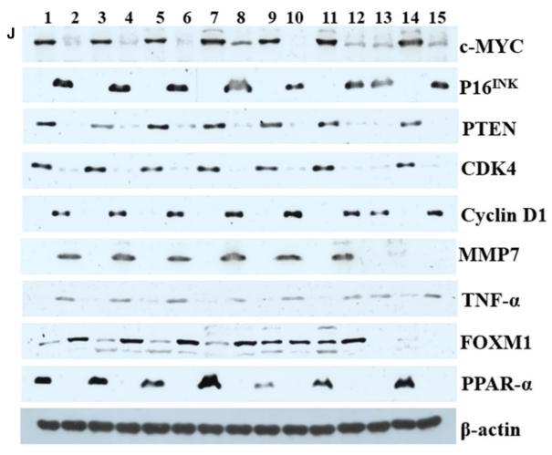
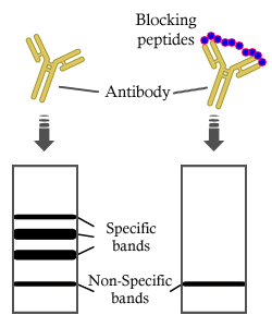
製品: PTEN Antibody
カタログ: AF6351
タンパク質の説明: Rabbit polyclonal antibody to PTEN
アプリケーション: WB IHC IF/ICC
Cited expt.: WB, IHC, IF/ICC
反応性: Human, Mouse, Rat, Monkey
予測: Pig, Bovine, Horse, Rabbit, Dog, Chicken, Xenopus
分子量: 54kDa(Observed); 47kD(Calculated).
ユニプロット: P60484
RRID: AB_2835156

類似製品を見る>>

   サイズ 価格 在庫状況
 100ul $280 在庫あり
 200ul $350 在庫あり

リードタイム: 当日配達

For pricing and ordering contact:
お問い合わせ先

製品説明

ソース:
Rabbit
アプリケーション:
WB 1:500-1:2000, IHC 1:50-1:200, IF/ICC 1:100-1:500
*The optimal dilutions should be determined by the end user. For optimal experimental results, antibody reuse is not recommended.
*Tips:

WB: For western blot detection of denatured protein samples. IHC: For immunohistochemical detection of paraffin sections (IHC-p) or frozen sections (IHC-f) of tissue samples. IF/ICC: For immunofluorescence detection of cell samples. ELISA(peptide): For ELISA detection of antigenic peptide.

反応性:
Human,Mouse,Rat,Monkey
予測:
Pig(100%), Bovine(100%), Horse(100%), Rabbit(100%), Dog(100%), Chicken(100%), Xenopus(100%)
クローナリティ:
Polyclonal
特異性:
PTEN Antibody detects endogenous levels of total PTEN.
RRID:
AB_2835156
引用形式: Affinity Biosciences Cat# AF6351, RRID:AB_2835156.
コンジュゲート:
Unconjugated.
精製:
The antiserum was purified by peptide affinity chromatography using SulfoLink™ Coupling Resin.
保存:
Rabbit IgG in phosphate buffered saline , pH 7.4, 150mM NaCl, 0.02% sodium azide and 50% glycerol. Store at -20 °C. Stable for 12 months from date of receipt.
別名:

折りたたみ/展開

10q23del; BZS; DEC; GLM2; MGC11227; MHAM; MMAC1; MMAC1 phosphatase and tensin homolog deleted on chromosome 10; Mutated in multiple advanced cancers 1; Phosphatase and tensin homolog; Phosphatase and tensin like protein; Phosphatidylinositol 3,4,5-trisphosphate 3-phosphatase and dual-specificity protein phosphatase PTEN; Pten; PTEN_HUMAN; PTEN1; TEP1;

免疫原

免疫原:

A synthesized peptide derived from human PTEN, corresponding to a region within C-terminal amino acids.

Uniprot:
遺伝子(ID):
発現特異性:
P60484 PTEN_HUMAN:

Expressed at a relatively high level in all adult tissues, including heart, brain, placenta, lung, liver, muscle, kidney and pancreas.

タンパク質の説明:
PTEN a phosphoinositide 3-phosphatase (PIP3) and a tumor suppressor implicated in a wide variety of human cancers. Dephosphorylates inositol phospholipids generated by the activation of the phosphoinositide 3 kinase (PI3K).
タンパク質配列:
MTAIIKEIVSRNKRRYQEDGFDLDLTYIYPNIIAMGFPAERLEGVYRNNIDDVVRFLDSKHKNHYKIYNLCAERHYDTAKFNCRVAQYPFEDHNPPQLELIKPFCEDLDQWLSEDDNHVAAIHCKAGKGRTGVMICAYLLHRGKFLKAQEALDFYGEVRTRDKKGVTIPSQRRYVYYYSYLLKNHLDYRPVALLFHKMMFETIPMFSGGTCNPQFVVCQLKVKIYSSNSGPTRREDKFMYFEFPQPLPVCGDIKVEFFHKQNKMLKKDKMFHFWVNTFFIPGPEETSEKVENGSLCDQEIDSICSIERADNDKEYLVLTLTKNDLDKANKDKANRYFSPNFKVKLYFTKTVEEPSNPEASSSTSVTPDVSDNEPDHYRYSDTTDSDPENEPFDEDQHTQITKV

種類予測

種類予測:

Score>80(red) has high confidence and is suggested to be used for WB detection. *The prediction model is mainly based on the alignment of immunogen sequences, the results are for reference only, not as the basis of quality assurance.

Species
Results
Score
Pig
100
Horse
100
Bovine
100
Dog
100
Xenopus
100
Chicken
100
Rabbit
100
Sheep
0
Zebrafish
0
Model Confidence:
High(score>80) Medium(80>score>50) Low(score<50) No confidence

研究背景

機能:

Tumor suppressor. Acts as a dual-specificity protein phosphatase, dephosphorylating tyrosine-, serine- and threonine-phosphorylated proteins. Also acts as a lipid phosphatase, removing the phosphate in the D3 position of the inositol ring from phosphatidylinositol 3,4,5-trisphosphate, phosphatidylinositol 3,4-diphosphate, phosphatidylinositol 3-phosphate and inositol 1,3,4,5-tetrakisphosphate with order of substrate preference in vitro PtdIns(3,4,5)P3 > PtdIns(3,4)P2 > PtdIns3P > Ins(1,3,4,5)P4. The lipid phosphatase activity is critical for its tumor suppressor function. Antagonizes the PI3K-AKT/PKB signaling pathway by dephosphorylating phosphoinositides and thereby modulating cell cycle progression and cell survival. The unphosphorylated form cooperates with AIP1 to suppress AKT1 activation. Dephosphorylates tyrosine-phosphorylated focal adhesion kinase and inhibits cell migration and integrin-mediated cell spreading and focal adhesion formation. Plays a role as a key modulator of the AKT-mTOR signaling pathway controlling the tempo of the process of newborn neurons integration during adult neurogenesis, including correct neuron positioning, dendritic development and synapse formation. May be a negative regulator of insulin signaling and glucose metabolism in adipose tissue. The nuclear monoubiquitinated form possesses greater apoptotic potential, whereas the cytoplasmic nonubiquitinated form induces less tumor suppressive ability. In motile cells, suppresses the formation of lateral pseudopods and thereby promotes cell polarization and directed movement.

Functional kinase, like isoform 1 it antagonizes the PI3K-AKT/PKB signaling pathway. Plays a role in mitochondrial energetic metabolism by promoting COX activity and ATP production, via collaboration with isoform 1 in increasing protein levels of PINK1.

PTMs:

Constitutively phosphorylated by CK2 under normal conditions. Phosphorylated in vitro by MAST1, MAST2, MAST3 and STK11. Phosphorylation results in an inhibited activity towards PIP3. Phosphorylation can both inhibit or promote PDZ-binding. Phosphorylation at Tyr-336 by FRK/PTK5 protects this protein from ubiquitin-mediated degradation probably by inhibiting its binding to NEDD4. Phosphorylation by ROCK1 is essential for its stability and activity. Phosphorylation by PLK3 promotes its stability and prevents its degradation by the proteasome.

Monoubiquitinated; monoubiquitination is increased in presence of retinoic acid. Deubiquitinated by USP7; leading to its nuclear exclusion. Monoubiquitination of one of either Lys-13 and Lys-289 amino acid is sufficient to modulate PTEN compartmentalization. Ubiquitinated by XIAP/BIRC4.

細胞の位置付け:

Cytoplasm. Nucleus. Nucleus>PML body.
Note: Monoubiquitinated form is nuclear. Nonubiquitinated form is cytoplasmic. Colocalized with PML and USP7 in PML nuclear bodies (PubMed:18716620). XIAP/BIRC4 promotes its nuclear localization (PubMed:19473982).

Secreted.
Note: May be secreted via a classical signal peptide and reenter into cells with the help of a poly-Arg motif.

Extracellular region or secreted Cytosol Plasma membrane Cytoskeleton Lysosome Endosome Peroxisome ER Golgi apparatus Nucleus Mitochondrion Manual annotation Automatic computational assertionSubcellular location
組織特異性:

Expressed at a relatively high level in all adult tissues, including heart, brain, placenta, lung, liver, muscle, kidney and pancreas.

タンパク質ファミリー:

The C2 domain binds phospholipid membranes in vitro in a Ca(2+)-independent manner; this binding is important for its tumor suppressor function.

Belongs to the PTEN phosphatase protein family.

研究領域

· Cellular Processes > Cell growth and death > p53 signaling pathway.   (View pathway)

· Cellular Processes > Transport and catabolism > Autophagy - animal.   (View pathway)

· Cellular Processes > Cell growth and death > Cellular senescence.   (View pathway)

· Cellular Processes > Cellular community - eukaryotes > Focal adhesion.   (View pathway)

· Environmental Information Processing > Signal transduction > FoxO signaling pathway.   (View pathway)

· Environmental Information Processing > Signal transduction > Phosphatidylinositol signaling system.

· Environmental Information Processing > Signal transduction > Sphingolipid signaling pathway.   (View pathway)

· Environmental Information Processing > Signal transduction > mTOR signaling pathway.   (View pathway)

· Environmental Information Processing > Signal transduction > PI3K-Akt signaling pathway.   (View pathway)

· Human Diseases > Drug resistance: Antineoplastic > EGFR tyrosine kinase inhibitor resistance.

· Human Diseases > Endocrine and metabolic diseases > Insulin resistance.

· Human Diseases > Infectious diseases: Viral > Hepatitis B.

· Human Diseases > Infectious diseases: Viral > Human papillomavirus infection.

· Human Diseases > Cancers: Overview > Pathways in cancer.   (View pathway)

· Human Diseases > Cancers: Overview > MicroRNAs in cancer.

· Human Diseases > Cancers: Specific types > Endometrial cancer.   (View pathway)

· Human Diseases > Cancers: Specific types > Glioma.   (View pathway)

· Human Diseases > Cancers: Specific types > Prostate cancer.   (View pathway)

· Human Diseases > Cancers: Specific types > Melanoma.   (View pathway)

· Human Diseases > Cancers: Specific types > Small cell lung cancer.   (View pathway)

· Human Diseases > Cancers: Specific types > Breast cancer.   (View pathway)

· Human Diseases > Cancers: Specific types > Hepatocellular carcinoma.   (View pathway)

· Human Diseases > Cancers: Overview > Central carbon metabolism in cancer.   (View pathway)

· Metabolism > Carbohydrate metabolism > Inositol phosphate metabolism.

参考文献

1). MiR‐19b‐3p accelerates bone loss after spinal cord injury by suppressing osteogenesis via regulating PTEN/Akt/mTOR signalling. JOURNAL OF CELLULAR AND MOLECULAR MEDICINE, 2021 (PubMed: 33332749) [IF=5.3]

Application: WB    Species: Human    Sample: HEK293T cells

FIGURE 3 MiR‐19b‐3p overexpression suppressed autophagy via binging to PTEN. (A) Dual‐Luciferase reporter assay was conducted to verify the targeted relationship between miR‐19b‐3p and 3’‐UTR of PTEN in HEK293T cells. Western blot for (B) PTEN, (C) p‐Akt, (D) p‐mTOR, (E) LC3II/I, (F) p62 and (G) Beclin‐1 in BMSC‐derived osteoblasts after transduction. Data were presented as means ± SD, and the statistical difference between two groups was determined by Student's t test. # P < .05 vs. LV‐Con. All the in vitro experiments were biologically repeated for three times (n = 3)

2). MiR-455-3p reduces apoptosis and alleviates degeneration of chondrocyte through regulating PI3K/AKT pathway. Life Sciences, 2020 (PubMed: 32343998) [IF=5.2]

Application: IHC    Species: Mice    Sample: BMSC

Fig. 2. Different gene expression profile between healthy cartilage and OA cartilage. A–D: miR-455-3p, PTEN, COL2A1, and MMP13 expression in healthy and OA cartilage by qRT-PCR, *P < 0.05, **P < 0.01, ***P < 0.001. E–F: Immunohistochemical results of Alcian, COL2A1, MMP13, PTEN, and pAKT in healthy and OA cartilage. (Scale: 400 μm).

Application: WB    Species: Mice    Sample: BMSC

Fig. 3. IL-1β/TNF-α down-regulates miR-455-3p and miR-455-3p inhibits the expression of PTEN. A: qPCR analyzing chondrocytes treated with IL-1β/TNF-α: IL-1β/TNF-α results in down-regulation of miR-455-3p and up-regulation of PTEN. B: The western blotting results of healthy cartilage and IL-1β treated cartilage. C–D: qRT-PCR analyzing overexpressing or inhibiting miR-455-3p: miR-455-3p inhibits PTEN and regulates related mRNA. E: western blotting analyzing the changes of related protein after transfecting mimic/inhibitor. *P < 0.05, **P < 0.01, ***P < 0.001.

3). Notch1 contributes to TNF-α-induced proliferation and migration of airway smooth muscle cells through regulation of the Hes1/PTEN axis. International Immunopharmacology, 2020 (PubMed: 32871474) [IF=4.8]

Application: WB    Species: Mice    Sample: ASM cells
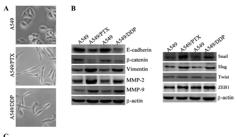
Fig. 5. Notch1 knockdown decreased Hes1 and increased PTEN expression in TNF-α-induced ASM cells. ASM cells were transfected with NC or Notch1 siRNA for 48 h and then stimulated with TNF-α for 24 h. (A, B) Protein expression of NICD was determined by western blot. (C) Relative Hes1 mRNA expression was detected by qPCR. (D, E) Protein expression of Hes1 was examined by western blot. (F, G) Protein expression of PTEN was measured by western blot. N = 3, **p < 0.01.

4). MicroRNA-181a regulates epithelial-mesenchymal transition by targeting PTEN in drug-resistant lung adenocarcinoma cells. INTERNATIONAL JOURNAL OF ONCOLOGY, 2015 (PubMed: 26323677) [IF=4.5]

Application: WB    Species: human    Sample: A549 cells
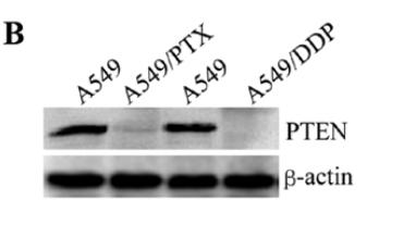
Figure 3. A549/DDP and A549/PTX cells showed molecular and morphological changes that were consistent with EMT. (A) microscopy at x200 magnification was used to assess cell morphology. The A549 cells (parental cells) had an epithelioid, rounded cobblestone appearance and there was limited formation of pseudopodia. A549/PTX and A549/DDP cells exhibited a spindle-shaped morphology and an increased formation of pseudopodia, indicating a loss of cell polarity. (B) E-cadherin, β-catenin, vimentin, MMP-2 and MMP-9 which are EMT-related proteins, were assessed in terms of expression levels. EMT-related transcription factors (Snail, Slug, Twist and ZEB1) were measured in A549/PTX and A549/DDP cells using western blot analysis. (C) The expression changes were confirmed at the mRNA level by qRT-PCR. Expression was standardized to the expression of GAPDH and normalized to 1.0 in the parental cells (compared with the parental A549 cells, means ± SEM, n=3, * P<0.05)

Application: WB    Species: human    Sample: lung adenocarcinoma cell

Figure 9.| miR-181a directly targets PTEN by binding to its 3'UTR. (A) The predicted miR-181a binding site within PTEN 3'UTR and its mutant version resulting from site mutagenesis are presented. Red indicates the nucleotides that were mutated to create a mismatch. (B) PTEN expression in lung adenocarcinoma cell lines was assessed by western blotting.

5). Mycobacterium tuberculosis ESAT6 modulates host innate immunity by downregulating miR-222-3p target PTEN. Biochimica et Biophysica Acta (BBA) - Molecular Basis of Disease, 2022 (PubMed: 34710568) [IF=4.2]

6). circIQCH sponges miR-145 to promote breast cancer progression by upregulating DNMT3A expression. Aging-US, 2020 (PubMed: 32756009) [IF=3.9]

Application: WB    Species: Mouse    Sample: breast cancer cell

Figure 5 circIQCH promotes breast cancer progression via circIQCH-miR-145-DNMT3A axis. (A) Predicted binding sites of miR-145 within the 3’-UTR of DNMT3A mRNA according to TargetScan. (B) The relative expression level of DNMT3A in breast cancer cell lines. (C) Luciferase reporter assay of SKBR3 and BT474 cells co-transfected with miR-145 mimics and the 3’-UTR of DNMT3A wild type or mutant luciferase reporter. The putative miRNA binding site of 3’-UTR of DNMT3A was mutated. (D) Expression of DNMT3A was decreased after transfection with miR-145 mimics. Expression of DNMT3A was increased after transfection with miR-145 inhibitors. (E) Enrichment of circIQCH, DNMT3A and miR-145 on Ago2 assessed by RIP assay. (F) Enrichment of Ago2 to circIQCH was decreased while DNMT3A was increased after knockdown of circIQCH. (G) Knockdown of circIQCH resulted in the reduction of DNMT3A expression which was reversed by miR-145 inhibitors. PTEN and BRCA1 was upregulated after silencing circIQCH. (H) Cell proliferation rate was detected by CCK-8 assay after exogenously expressing DNMT3A or inhibiting miR-145 in circIQCH silencing SKBR3 and BT474 cells. (I) Cell migration ability was validated by transwell assay after exogenously expressing DNMT3A or inhibiting miR-145 in circIQCH silencing SKBR3 cells. *P<0.05; **P<0.01.

7). Diosmin mitigates high glucose-induced endoplasmic reticulum stress through PI3K/AKT pathway in HK-2 cells. BMC Complementary Medicine and Therapies, 2022 (PubMed: 35477428) [IF=3.9]

Application: WB    Species: human    Sample: HK-2 cells

Fig. 7| The efect of diosmin on PI3K/AKT/PTEN signal transduction in high glucose-mediated HK-2 cells. A-D. The efect of diosmin on phosphatidylinositol 3-kinase/protein kinase-B/ phosphatase and tensin homolog deleted on chromosome 10 pathway-related markers was detected by western blot. β-actin was used as a housekeeping gene. The results were expressed as the mean±standard deviation (SD) of three independent experiments.

8). HDAC6, modulated by miR-206, promotes endometrial cancer progression through the PTEN/AKT/mTOR pathway. Scientific Reports, 2020 (PubMed: 32107418) [IF=3.8]

Application: WB    Species: Human    Sample: EC cells

Figure 6 Pathways modulated by HDAC6 in EC cells. (A) Western blotting showed the levels of differentially expressed proteins in Ishikawa cells following transfection with generated lentiviruses. (B) Putative working model depicting the role of miR-206/HDAC6 in EC progression. *P < 0.05, **P < 0.01, ***P < 0.001.

9). Lentinan induces apoptosis of mouse hepatocellular carcinoma cells through the EGR1/PTEN/AKT signaling axis. Oncology Reports, 2023 (PubMed: 37264970) [IF=3.8]

Application: WB    Species: Mouse    Sample: Hepa1-6 cells

Figure 2. Effects of LNT on the EGR1/PTEN/AKT axis in Hepa1-6 cells. (A) Protein expression levels of EGR1, PTEN, p-Akt and phosphorylation of Akt after treatment with a gradient of LNT concentrations as detected by WB. All data for protein expression were normalized using β-actin as a loading reference. (B) Immunofluorescence co-staining was used to assess the localization and expression of EGR1 and PTEN in Hepa1-6 cells treated with a gradient of LNT concentrations. Images were observed at ×630 magnification. Scale bars, 50 µm (white). (C) WB was used to detect the expression of EGR1 in the nuclear and cytoplasmic fractions following treatment with a gradient of LNT concentrations. (D) WB was used to detect the expression of EGR1, PTEN, p-Akt, and phosphorylation of Akt after EGR1 overexpression. *P

Application: IF/ICC    Species: Mouse    Sample: Hepa1-6 cells

Figure 2. Effects of LNT on the EGR1/PTEN/AKT axis in Hepa1-6 cells. (A) Protein expression levels of EGR1, PTEN, p-Akt and phosphorylation of Akt after treatment with a gradient of LNT concentrations as detected by WB. All data for protein expression were normalized using β-actin as a loading reference. (B) Immunofluorescence co-staining was used to assess the localization and expression of EGR1 and PTEN in Hepa1-6 cells treated with a gradient of LNT concentrations. Images were observed at ×630 magnification. Scale bars, 50 µm (white). (C) WB was used to detect the expression of EGR1 in the nuclear and cytoplasmic fractions following treatment with a gradient of LNT concentrations. (D) WB was used to detect the expression of EGR1, PTEN, p-Akt, and phosphorylation of Akt after EGR1 overexpression. *P

10). Regulation of the PTEN/PI3K/AKT pathway in RCC using the active compounds of natural products in vitro. Molecular Medicine Reports, 2021 (PubMed: 34490473) [IF=3.4]

Application: WB    Species: human    Sample: 786‑O and OS‑RC‑2 cells

Figure 5.| Naringenin inhibits renal cell carcinoma progression via activation of the PTEN/PI3K/AKT signaling pathway. Western blot analysis of PTEN,mTOR, p‑mTOR, PI3K, AKT and p‑AKT expression in 786‑O and OS‑RC‑2 cells. Data are presented as the mean ± SD. *P<0.05, **P<0.01 vs. Abs. Abs, abso‑lute ethanol; p‑, phosphorylated.

Application: WB    Species: Human    Sample: 786-O and OS-RC-2 cells

Figure 5. Naringenin inhibits renal cell carcinoma progression via activation of the PTEN/PI3K/AKT signaling pathway. Western blot analysis of PTEN, mTOR, p-mTOR, PI3K, AKT and p-AKT expression in 786-O and OS-RC-2 cells. Data are presented as the mean ± SD. *P<0.05, **P<0.01 vs. Abs. Abs, absolute ethanol; p-, phosphorylated.

もっと読みます

Restrictive clause


Affinity Biosciences tests all products strictly. Citations are provided as a resource for additional applications that have not been validated by Affinity Biosciences. Please choose the appropriate format for each application and consult Materials and Methods sections for additional details about the use of any product in these publications.

For Research Use Only.
Not for use in diagnostic or therapeutic procedures. Not for resale. Not for distribution without written consent. Affinity Biosciences will not be held responsible for patent infringement or other violations that may occur with the use of our products. Affinity Biosciences, Affinity Biosciences Logo and all other trademarks are the property of Affinity Biosciences LTD.