製品: Synapsin I Antibody
カタログ: AF6201
タンパク質の説明: Rabbit polyclonal antibody to Synapsin I
アプリケーション: WB IHC IF/ICC
Cited expt.: WB, IF/ICC
反応性: Human, Mouse, Rat
予測: Pig, Zebrafish, Bovine, Sheep, Rabbit, Xenopus
分子量: 77kDa; 74kD(Calculated).
ユニプロット: P17600
RRID: AB_2835082

類似製品を見る>>

   サイズ 価格 在庫状況
 100ul $280 在庫あり
 200ul $350 在庫あり

リードタイム: 当日配達

For pricing and ordering contact:
お問い合わせ先

製品説明

ソース:
Rabbit
アプリケーション:
WB 1:500-1:2000, IF/ICC 1:100-1:500, IHC 1:50-1:200
*The optimal dilutions should be determined by the end user. For optimal experimental results, antibody reuse is not recommended.
*Tips:

WB: For western blot detection of denatured protein samples. IHC: For immunohistochemical detection of paraffin sections (IHC-p) or frozen sections (IHC-f) of tissue samples. IF/ICC: For immunofluorescence detection of cell samples. ELISA(peptide): For ELISA detection of antigenic peptide.

反応性:
Human,Mouse,Rat
予測:
Pig(100%), Zebrafish(100%), Bovine(100%), Sheep(100%), Rabbit(100%), Xenopus(100%)
クローナリティ:
Polyclonal
特異性:
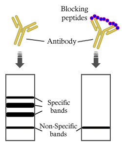
Synapsin I Antibody detects endogenous levels of total Synapsin I.
RRID:
AB_2835082
引用形式: Affinity Biosciences Cat# AF6201, RRID:AB_2835082.
コンジュゲート:
Unconjugated.
精製:
The antiserum was purified by peptide affinity chromatography using SulfoLink™ Coupling Resin (Thermo Fisher Scientific).
保存:
Rabbit IgG in phosphate buffered saline , pH 7.4, 150mM NaCl, 0.02% sodium azide and 50% glycerol. Store at -20 °C. Stable for 12 months from date of receipt.
別名:

折りたたみ/展開

Brain protein 4.1; SYN 1; SYN 1a; SYN 1b; SYN I; SYN1; SYN1_HUMAN; SYN1a; SYN1b; Synapsin 1; Synapsin I; Synapsin-1; Synapsin1; SynapsinI; SYNI;

免疫原

免疫原:

A synthesized peptide derived from human Synapsin I, corresponding to a region within N-terminal amino acids.

Uniprot:
遺伝子(ID):
タンパク質の説明:
This gene is a member of the synapsin gene family. Synapsins encode neuronal phosphoproteins which associate with the cytoplasmic surface of synaptic vesicles. Family members are characterized by common protein domains, and they are implicated in synaptogenesis and the modulation of neurotransmitter release, suggesting a potential role in several neuropsychiatric diseases.
タンパク質配列:
MNYLRRRLSDSNFMANLPNGYMTDLQRPQPPPPPPGAHSPGATPGPGTATAERSSGVAPAASPAAPSPGSSGGGGFFSSLSNAVKQTTAAAAATFSEQVGGGSGGAGRGGAASRVLLVIDEPHTDWAKYFKGKKIHGEIDIKVEQAEFSDLNLVAHANGGFSVDMEVLRNGVKVVRSLKPDFVLIRQHAFSMARNGDYRSLVIGLQYAGIPSVNSLHSVYNFCDKPWVFAQMVRLHKKLGTEEFPLIDQTFYPNHKEMLSSTTYPVVVKMGHAHSGMGKVKVDNQHDFQDIASVVALTKTYATAEPFIDAKYDVRVQKIGQNYKAYMRTSVSGNWKTNTGSAMLEQIAMSDRYKLWVDTCSEIFGGLDICAVEALHGKDGRDHIIEVVGSSMPLIGDHQDEDKQLIVELVVNKMAQALPRQRQRDASPGRGSHGQTPSPGALPLGRQTSQQPAGPPAQQRPPPQGGPPQPGPGPQRQGPPLQQRPPPQGQQHLSGLGPPAGSPLPQRLPSPTSAPQQPASQAAPPTQGQGRQSRPVAGGPGAPPAARPPASPSPQRQAGPPQATRQTSVSGPAPPKASGAPPGGQQRQGPPQKPPGPAGPTRQASQAGPVPRTGPPTTQQPRPSGPGPAGRPKPQLAQKPSQDVPPPATAAAGGPPHPQLNKSQSLTNAFNLPEPAPPRPSLSQDEVKAETIRSLRKSFASLFSD

種類予測

種類予測:

Score>80(red) has high confidence and is suggested to be used for WB detection. *The prediction model is mainly based on the alignment of immunogen sequences, the results are for reference only, not as the basis of quality assurance.

Species
Results
Score
Pig
100
Bovine
100
Sheep
100
Xenopus
100
Zebrafish
100
Rabbit
100
Horse
0
Dog
0
Chicken
0
Model Confidence:
High(score>80) Medium(80>score>50) Low(score<50) No confidence

研究背景

機能:

Neuronal phosphoprotein that coats synaptic vesicles, binds to the cytoskeleton, and is believed to function in the regulation of neurotransmitter release. The complex formed with NOS1 and CAPON proteins is necessary for specific nitric-oxid functions at a presynaptic level.

PTMs:

Substrate of at least four different protein kinases. It is probable that phosphorylation plays a role in the regulation of synapsin-1 in the nerve terminal.

Phosphorylation at Ser-9 dissociates synapsins from synaptic vesicles.

細胞の位置付け:

Cell junction>Synapse. Golgi apparatus.

Extracellular region or secreted Cytosol Plasma membrane Cytoskeleton Lysosome Endosome Peroxisome ER Golgi apparatus Nucleus Mitochondrion Manual annotation Automatic computational assertionSubcellular location
タンパク質ファミリー:

The A region binds phospholipids with a preference for negatively charged species.

Belongs to the synapsin family.

参考文献

1). The brain-protective mechanism of fecal microbiota transplantation from young donor mice in the natural aging process via exosome, gut microbiota, and metabolomics analyses. Pharmacological research, 2024 (PubMed: 39053865) [IF=9.1]

Application: WB    Species: Mouse    Sample:

Fig. 3. The cerebral protection effects of FMT in mice against age-associated proteins expression levels. (A, B) Quantified protein levels of the cell cycle arrest related proteins of p53, p21, p16/p14, and Rb in mice hippocampus tissues. Data are shown as the mean ± SD, n = 3 per group. Significance was determined by one-way ANOVA followed by the Dunnett T3 test for comparison of multiple groups, *P < 0.05, **P < 0.01, and ***P < 0.001. (C, D) Quantified protein levels of the DNA damage-related protein ATM, and the cognitive-related proteins synapsin I, synaptophysin and PSD95 in mice hippocampus tissues. Data are shown as the mean ± SD, n = 3 per group. Significance was determined by one-way ANOVA followed by the Dunnett T3 test for comparison of multiple groups, *P < 0.05, **P < 0.01, and ***P < 0.001. (E, F) Quantified protein levels of the cell senescence-related proteins CREB, p-CREB, ERK, p-ERK, AKT, p-AKT in mice hippocampus tissues. Data are shown as the mean ± SD, n = 3 per group. Significance was determined by one-way ANOVA followed by the Dunnett T3 test for comparison of multiple groups, *P < 0.05, **P < 0.01, and ***P < 0.001. (G, H) Quantified protein levels of the c-H2AX and TP53BP1 proteins in bone marrow mesenchymal stem cells. Data are shown as the mean ± SD, n = 3 per group. Significance was determined by one-way ANOVA followed by the Dunnett T3 test for comparison of multiple groups, *P < 0.05, **P < 0.01, and ***P < 0.001. (I, J) Quantified protein levels of the β-galactosidase, c-H2AX, and TP53BP1 proteins in in mice hippocampus tissues. Data are shown as the mean ± SD, n = 3 per group. Significance was determined by one-way ANOVA followed by the Dunnett T3 test for comparison of multiple groups, *P < 0.05, **P < 0.01, and ***P < 0.001.

2). Repetitive Trans-spinal Magnetic Stimulation Suppresses Microglia to Engulf Synapse and Promotes Nerve Repairment via cGAS-STING Signaling Pathway after Spinal Cord Injury. International journal of biological sciences, 2025 (PubMed: 41208875) [IF=8.2]

Application: WB    Species: Rat    Sample:

Figure 3. rTSMS enhances synapse formation and neuronal activity in rats with SCI. (A-B) Representative images of western blotting and quantitative analysis of PSD95, SYN and NF200 in the injured spinal cord at 21 dpi; n = 3. (C-D) Representative images of immunostaining and statistical analysis of NF200 (green), SYN (red) and DAPI (blue) in the injured spinal cord at 21 dpi; n = 5. (E) Z-stack image of NF200/SYN in the rTSMS group, slice thickness = 10 um, number of layers = 10. (F-G) Representative images of immunostaining and statistical analysis of CGRP (green), SYN (red) and DAPI (blue) in the injured spinal cord at 21 dpi, n = 5. (H-I) Representative images of immunostaining and statistical analysis of TH (green), SYN (red) and DAPI (blue) in the injured spinal cord at 21 dpi, n = 5. (J-K) Z-stack images of CGRP/SYN and TH/SYN in the rTSMS group, slice thickness = 10 um, number of layers = 10. (L) A schematic illustration showing the M1 and S1 regions in the brain. The image was generated with BioRender.com. (M) Representative immunostaining images of NEUN (green), cFOS (red) and DAPI (blue) in the S1 region at 21 dpi. (N) Statistical analysis of cFOS intensity in the M1 and S1 regions at 21 dpi, n = 5. *P < 0.05, **P < 0.01, ***P < 0.001.

Application: IF/ICC    Species: Rat    Sample:
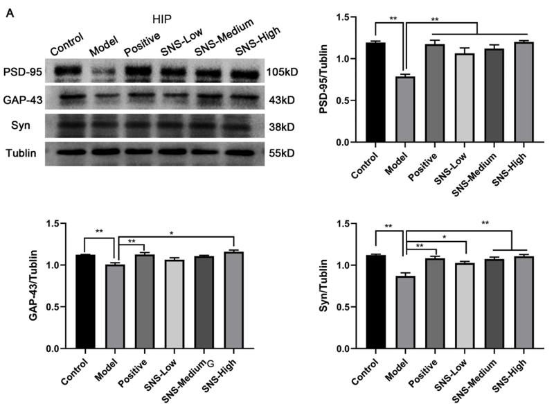
Figure 3. rTSMS enhances synapse formation and neuronal activity in rats with SCI. (A-B) Representative images of western blotting and quantitative analysis of PSD95, SYN and NF200 in the injured spinal cord at 21 dpi; n = 3. (C-D) Representative images of immunostaining and statistical analysis of NF200 (green), SYN (red) and DAPI (blue) in the injured spinal cord at 21 dpi; n = 5. (E) Z-stack image of NF200/SYN in the rTSMS group, slice thickness = 10 um, number of layers = 10. (F-G) Representative images of immunostaining and statistical analysis of CGRP (green), SYN (red) and DAPI (blue) in the injured spinal cord at 21 dpi, n = 5. (H-I) Representative images of immunostaining and statistical analysis of TH (green), SYN (red) and DAPI (blue) in the injured spinal cord at 21 dpi, n = 5. (J-K) Z-stack images of CGRP/SYN and TH/SYN in the rTSMS group, slice thickness = 10 um, number of layers = 10. (L) A schematic illustration showing the M1 and S1 regions in the brain. The image was generated with BioRender.com. (M) Representative immunostaining images of NEUN (green), cFOS (red) and DAPI (blue) in the S1 region at 21 dpi. (N) Statistical analysis of cFOS intensity in the M1 and S1 regions at 21 dpi, n = 5. *P < 0.05, **P < 0.01, ***P < 0.001.

3). SiNiSan ameliorates depression-like behavior in rats by enhancing synaptic plasticity via the CaSR-PKC-ERK signaling pathway. BIOMEDICINE & PHARMACOTHERAPY, 2020 (PubMed: 31958763) [IF=6.9]

Application: WB    Species: rat    Sample: HIP
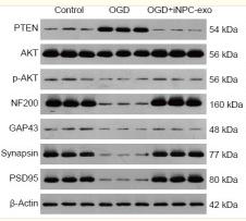
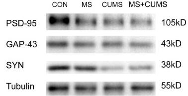
Fig. 5. |Effects of SNS on synaptic-associated protein in the HIP and PFC of stressed rats. Representative immunoblots for PSD-95, GAP-43, Syn, and Tublin in the HIP(A) and PFC(B) regions. (A) Results of relative protein levels of PSD-95, GAP-43, and Syn in the HIP of rats of each group.

4). Exosomes derived from human induced pluripotent stem cell-derived neural progenitor cells protect neuronal function under ischemic conditions. Neural Regeneration Research, 2021 (PubMed: 33642395) [IF=5.9]

Application: WB    Species: Rat    Sample: neural progenitor cells

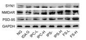
Figure 3 Effect of iPSC-NPC-derived exosomes (OGD+iNPC-exo) on expression of the PTEN/AKT signaling pathway and of synaptic plasticity-related proteins in OGD induced neurons. (A–C) mRNA expression (A) and protein expression (B, C) of the PTEN/AKT signaling pathway and of synaptic plasticity-related proteins (NF200, GAP-43, Synapsin, and PSD95) analyzed by polymerase chain reaction and western blot assay. The mRNA expression is described by the optical density ratio relative to the control group. Protein expression was described by the optical density ratio relative to β-actin. Data are presented as mean ± SD. ***P < 0.001, vs. control group; ###P < 0.001 (one-way analysis of variance with post hoc Bonferroni test). All experiments were repeated three times. GAP43: growth associated protein 43; iPSC-NPCs: induced pluripotent stem cells-derived neural progenitor cells; NF200: neurofilament 200; OGD: oxygen-glucose deprivation; p-Akt: phosphor-Akt; PSD95: postsynaptic density protein 95; PTEN: phosphatase and tensin homolog deleted on chromosome ten.

5). Neuron-glioma synaptic transmission amplified by free 19S proteasome-mediated AMPAR deubiquitination promotes tumor progression. Neurotherapeutics : the journal of the American Society for Experimental NeuroTherapeutics, 2026 (PubMed: 41667338) [IF=5.6]

6). Research on the anti-aging mechanisms of Panax ginseng extract in mice: a gut microbiome and metabolomics approach. Frontiers in pharmacology, 2024 (PubMed: 38966558) [IF=5.6]

Application: WB    Species: Mouse    Sample:
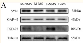
Figure 3. Protective effects of ginseng and FMT in mice against age-associated protein expression levels. (A,B) Quantified protein levels of cell cycle arrest-related proteins of p53, p21, p16/p14, and Rb in mouse hippocampus tissues. Data are shown as the mean ± SD, n = 3 per group. Significance was determined by one-way ANOVA, followed by the LSD post hoc test or Dunnett’s T3 test for the comparison of multiple groups; *p < 0.05, **p < 0.01, and ***p < 0.001. (C,D) Quantified protein levels of the DNA damage-related protein ATM and the cognitive-related proteins synapsin I, synaptophysin, and PSD95 in mouse hippocampus tissues. Data are shown as the mean ± SD, n = 3 per group. Significance was determined by one-way ANOVA, followed by the LSD post hoc test or Dunnett’s T3 test for the comparison of multiple groups; *p < 0.05, **p < 0.01, and ***p < 0.001. (E,F) Quantified protein levels of the cell senescence-related proteins CREB, p-CREB, ERK, p-ERK, AKT, and p-AKT in mouse hippocampus tissues. Data are shown as the mean ± SD, n = 3 per group. Significance was determined by one-way ANOVA, followed by the LSD post hoc test or Dunnett’s T3 test for the comparison of multiple groups; *p < 0.05, **p < 0.01, and ***p < 0.001. (G,H) Quantified levels of the c-H2AX and TP53BP1 proteins in bone marrow mesenchymal stem cells. Data are shown as the mean ± SD, n = 3 per group. Significance was determined by one-way ANOVA, followed by the LSD post hoc test or Dunnett’s T3 test for the comparison of multiple groups; *p < 0.05, **p < 0.01, and ***p < 0.001.

7). Early-Life Stress Induces Depression-Like Behavior and Synaptic-Plasticity Changes in a Maternal Separation Rat Model: Gender Difference and Metabolomics Study. Frontiers in Pharmacology, 2020 (PubMed: 32174832) [IF=5.6]

Application: WB    Species: Rat    Sample: hippocampus

Figure 5 MS reduces the expression of synaptic-plasticity protein. (A) The bands of synaptic-plasticity proteins of SYN, PSD-95, and GAP-43 in the hippocampus by WB. Statistical results indicate the relative protein levels expressed by SYN, GAP-43, and PSD-95. (B) The bands of synaptic-plasticity proteins of SYN, PSD-95, and GAP-43 in cortex by WB. Statistical results indicate the relative protein levels expressed by SYN, GAP-43, and PSD-95. Statistical analyses are performed by two-way ANOVA followed by t-test. Data are presented as mean ± SEM, *p < 0.05, **p < 0.01, n = 3 per group.

8). Ginsenoside Rg1 alleviates chronic inflammation-induced neuronal ferroptosis and cognitive impairments via regulation of AIM2 - Nrf2 signaling pathway. Journal of ethnopharmacology, 2024 (PubMed: 38641079) [IF=4.8]

9). Li, P HY-021068 alleviates cerebral ischemia-reperfusion injury by inhibiting NLRP1 inflammasome and restoring autophagy function in mice. Experimental neurology, 2024 (PubMed: 37884189) [IF=4.6]

10). ZuoGui Pill Ameliorates Alzheimer's Disease-Like Pathology in 3xTg-AD Mice by Targeting Aβ Production, Tau Phosphorylation, Synaptic Loss, and Neuroinflammation. Molecular neurobiology, 2025 (PubMed: 41261326) [IF=4.6]

Application: WB    Species: Mouse    Sample:

Fig. 4 ZGP reduces synaptic loss and neuropathological alterations in 3xTg-AD mice. A Representative NeuN IHC in the CA1 region (scale bar, 50 μm). B Quantification of NeuN-positive area in A. C Western blot analysis of hippocampal Syn and PSD-95 expression levels. D Quantification of protein expression levels from C. E Representative HE and Nissl staining in the CA1 region (scale bar, 50 μm). F Representative Golgi staining of hippocampal neurons. G Quantification of dendritic spine density per 10 μm from F. Data are presented as mean ± SEM (n = 5 per group for IHC and WB; n = 10 per group for Golgi staining). Shapiro–Wilk and Bartlett’s tests were used to assess normality and variance homogeneity. All data were analyzed using one-way ANOVA with Tukey’s post hoc test. *P 

もっと読みます

Restrictive clause


Affinity Biosciences tests all products strictly. Citations are provided as a resource for additional applications that have not been validated by Affinity Biosciences. Please choose the appropriate format for each application and consult Materials and Methods sections for additional details about the use of any product in these publications.

For Research Use Only.
Not for use in diagnostic or therapeutic procedures. Not for resale. Not for distribution without written consent. Affinity Biosciences will not be held responsible for patent infringement or other violations that may occur with the use of our products. Affinity Biosciences, Affinity Biosciences Logo and all other trademarks are the property of Affinity Biosciences LTD.