製品: Phospho-FUNDC1 (Ser17) antibody
カタログ: AF0001
タンパク質の説明: Rabbit polyclonal antibody to Phospho-FUNDC1 (Ser17)
アプリケーション: WB
Cited expt.: WB
反応性: Human
予測: Pig, Bovine, Horse, Sheep, Rabbit, Dog, Chicken
分子量: 17KD; 17kD(Calculated).
ユニプロット: Q8IVP5
RRID: AB_2846773

類似製品を見る>>

   サイズ 価格 在庫状況
 100ul $280 在庫あり
 200ul $350 在庫あり

リードタイム: 当日配達

For pricing and ordering contact:
お問い合わせ先

製品説明

ソース:
Rabbit
アプリケーション:
WB 1:500-1:2000
*The optimal dilutions should be determined by the end user. For optimal experimental results, antibody reuse is not recommended.
*Tips:

WB: For western blot detection of denatured protein samples. IHC: For immunohistochemical detection of paraffin sections (IHC-p) or frozen sections (IHC-f) of tissue samples. IF/ICC: For immunofluorescence detection of cell samples. ELISA(peptide): For ELISA detection of antigenic peptide.

反応性:
Human
予測:
Pig(100%), Bovine(100%), Horse(100%), Sheep(100%), Rabbit(100%), Dog(88%), Chicken(100%)
クローナリティ:
Polyclonal
特異性:
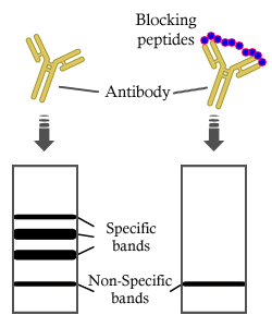
Phospho-FUNDC1 (Ser17) antibody detects endogenous levels of FUNDC1 only when phosphorylated at Serine 17.
RRID:
AB_2846773
引用形式: Affinity Biosciences Cat# AF0001, RRID:AB_2846773.
コンジュゲート:
Unconjugated.
精製:
The antibody is from purified rabbit serum by affinity purification via sequential chromatography on phospho-peptide and non-phospho-peptide affinity columns.
保存:
Rabbit IgG in phosphate buffered saline , pH 7.4, 150mM NaCl, 0.02% sodium azide and 50% glycerol. Store at -20 °C. Stable for 12 months from date of receipt.
別名:

折りたたみ/展開

FUN14 domain containing 1; FUN14 domain containing protein 1; FUN14 domain-containing protein 1; FUND1_HUMAN; fundc1;

免疫原

免疫原:

A synthesized peptide derived from human FUNDC1 around the phosphorylation site of Serine 17.

Uniprot:
遺伝子(ID):
発現特異性:
Q8IVP5 FUND1_HUMAN:

Widely expressed.

タンパク質配列:
MATRNPPPQDYESDDDSYEVLDLTEYARRHQWWNRVFGHSSGPMVEKYSVATQIVMGGVTGWCAGFLFQKVGKLAATAVGGGFLLLQIASHSGYVQIDWKRVEKDVNKAKRQIKKRANKAAPEINNLIEEATEFIKQNIVISSGFVGGFLLGLAS

種類予測

種類予測:

Score>80(red) has high confidence and is suggested to be used for WB detection. *The prediction model is mainly based on the alignment of immunogen sequences, the results are for reference only, not as the basis of quality assurance.

Species
Results
Score
Pig
100
Horse
100
Bovine
100
Sheep
100
Chicken
100
Rabbit
100
Dog
88
Xenopus
0
Zebrafish
0
Model Confidence:
High(score>80) Medium(80>score>50) Low(score<50) No confidence

研究背景

機能:

Acts as an activator of hypoxia-induced mitophagy, an important mechanism for mitochondrial quality control.

PTMs:

Phosphorylation at Tyr-18 by SRC inhibits activation of mitophagy. Following hypoxia, dephosphorylated at Tyr-18, leading to interaction with MAP1 LC3 family proteins and triggering mitophagy.

細胞の位置付け:

Mitochondrion outer membrane>Multi-pass membrane protein.

Extracellular region or secreted Cytosol Plasma membrane Cytoskeleton Lysosome Endosome Peroxisome ER Golgi apparatus Nucleus Mitochondrion Manual annotation Automatic computational assertionSubcellular location
組織特異性:

Widely expressed.

タンパク質ファミリー:

The YXXL motif mediates the interaction with MAP1 LC3 family proteins MAP1LC3A, MAP1LC3B and GABARAP.

Belongs to the FUN14 family.

参考文献

1). Inhibiting CD36 palmitoylation improves cardiac function post-infarction by regulating lipid metabolic homeostasis and autophagy. Nature communications, 2025 (PubMed: 40675975) [IF=16.6]

Application: WB    Species: Mouse    Sample:

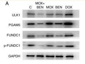
Fig. 9: Insights into the role of CD36 palmitoylation in cardiomyocyte autophagy through PGAM5. a Western blotting analysis of LC3II and SQSTM1 protein levels in myocardium from WT-CD36 and AA-SS-CD36 mice. n = 7. b Western blotting analysis of LC3II and SQSTM1 protein levels in cardiomyocytes transfected with WT-CD36 or AA-SS-CD36 for 48 h. n = 10. c Overlap between CD36 binding proteins, as assessed by CoIP-MS analysis and proteins in the Autophagy Database (http://autophagy.info/). d Endogenous CD36 immunoprecipitation followed by anti-CD36 and anti-PGAM5 western blot analysis of cardiomyocytes (NMVCs). n = 4. e Quantification of Cyto-ID staining in cardiomyocytes (NMVCs) transfected with WT-CD36 or AA-SS-CD36 for 48 h, and then treated with 500 nM rapamycin (Rapa) and 60 µM CQ, the combination for 16 h. n = 6 per group. f–h Western blotting analysis of the mitochondria expression of CD36 and PGAM5 in cardiomyocytes (NMVCs) transfected with WT-CD36 or AA-SS-CD36. n = 7 per group. i Relative PGAM5 mRNA levels in cardiomyocytes transfected with WT-CD36 or AA-SS-CD36 for 48 h. n = 5. j Time course of PGAM5 protein stability in cardiomyocytes (NMVCs) transfected with WT-CD36 or AA-SS-CD36 after treatment with CHX (10 µg/ml). n = 6. k Ubiquitination of exogenous PGAM5 in cardiomyocytes (NMVCs) transfected with WT-CD36 or AA-SS-CD36. Data are representative of six independent experiments. l The mitochondria expression of phosphorylation Fundc1 and total Fundc1 protein in cardiomyocytes (NMVCs) transfected with WT-CD36 or AA-SS-CD36. n = 6 for phosphorylation Fundc1; n = 7 for total Fundc1. Data are presented as means ± SEM. Statistical significance was assessed by Kruskal-Wallis, followed by false discovery rate [FDR] method of Benjamini and Hochberg test (a), two-tailed unpaired Student t test (b, d, g, i and l), two-way ANOVA, followed by Tukey post hoc multicomparisons test (e, j) and two-tailed Mann-Whitney U test (h). Source data are provided as a Source Data file.

2). TrxR2 Lactylation Facilitates Mitochondrial Protection and Endothelial Ferroptosis Resistance in Diabetic Cardiomyopathy. Advanced science (Weinheim, Baden-Wurttemberg, Germany), 2026 (PubMed: 41704008) [IF=15.1]

3). DDX21 mediates co-transcriptional RNA m6A modification to promote transcription termination and genome stability. Molecular cell, 2024 (PubMed: 38569554) [IF=14.5]

4). Pharmacological inhibition of Septins with Forchlorfenuron attenuates thrombus formation in experimental thrombotic mice models with modulating multiple signaling pathways in platelets. Journal of advanced research, 2024 (PubMed: 39111626) [IF=11.4]

5). c-FLIP Protects Cardiac Microcirculation in Sepsis-Induced Myocardial Dysfunction Via FUNDC1-Mediated Regulation of Mitochondrial Autophagy. JACC. Basic to translational science, 2025 (PubMed: 40372306) [IF=8.4]

Application: WB    Species: rat    Sample:

Figure 3. Construction of In Vitro Model and Observation of Mitochondrial Autophagy and Expression Changes of c-FLIP, JNK, and FUNDC1 (A and B) Optical microscopy of cardiac microvascular endothelial cell (CMEC) morphology and CCK-8 assay for cell proliferation after 12 hours of different lipopolysaccharide (LPS) concentrations. (C and D) Optical microscopy of CMECs morphology and CCK-8 assay for cell proliferation at various times after 10 μg/mL LPS treatment. (E and F) c-FLIP expression at various times after 10 μg/mL LPS treatment. (G) Transmission electron microscopy (×12,000) of CMECs microstructure to assess mitochondrial damage and autophagy (representative images). (H to L) Western blot of mitochondrial autophagy-related proteins. (M-Q) Expression of c-FLIP, total c-Jun N-terminal kinase (t-JNK), phosphorylated JNK (p-JNK), total Fun14 domain-containing protein 1 (t-FUNDC1), phosphorylated FUNDC1 (p-FUNDC1) proteins, and FUNDC1 mRNA. Note: n = 3/group; 3 technical replicates; mean ± SEM; Student's t-test (N-Q), analysis of variance with Tukey’s post hoc test (B, D, F, and I to L). ∗P < 0.05 vs Ctrl (control) group, ∗∗P < 0.01 vs Ctrl group, ∗∗∗P < 0.001 vs Ctrl group. Abbreviations as in Figure 1.

6). TBK1 Facilitates GLUT1-Dependent Glucose Consumption by suppressing mTORC1 Signaling in Colorectal Cancer Progression. International Journal of Biological Sciences, 2023 (PubMed: 35637944) [IF=8.2]

7). FUNDC1-mediated mitophagy triggered by mitochondrial ROS is partially involved in 1-nitropyrene-evoked placental progesterone synthesis inhibition and intrauterine growth retardation in mice. The Science of the total environment, 2024 (PubMed: 37951264) [IF=8.2]

8). NTRK1 knockdown induces mouse cognitive impairment and hippocampal neuronal damage through mitophagy suppression via inactivating the AMPK/ULK1/FUNDC1 pathway. Cell death discovery, 2023 (PubMed: 37907480) [IF=7.0]

Application: WB    Species: Mouse    Sample:

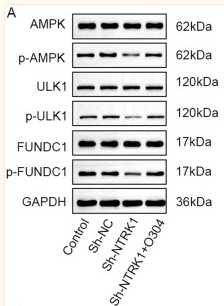
Fig. 6 NTRK1 silencing might suppress mitophagy in mouse neurons through the inactivation of the AMPK/ULK1/FUNDC1 pathway. A, B WB revealed that O304 treatment abrogated the suppression effect of NTRK1 knockdown on the activity of the AMPK/ULK1/FUNDC1 pathway in mouse neurons. n = 3. C, D WB also indicated that O304 treatment counteracted the suppression effect of NTRK1 silencing on mitophagy in mouse neurons. n = 3. E, F The results of TOMM20/LC3-II fluorescence staining revealed that the O304 treatment reversed the suppression effect of NTRK1 knockdown on the expression of Parkin in mouse neuronmitochondria. n = 3.

9). Xanthohumol Regulates Mitophagy in Osteosarcoma Cells via AMPK-ULK1-FUNDC1 Signaling Pathway. Phytotherapy research : PTR, 2025 (PubMed: 40190139) [IF=6.1]

10). Hypoxia‑induced mitophagy regulates proliferation, migration and odontoblastic differentiation of human dental pulp cells through FUN14 domain‑containing 1. International Journal of Molecular Medicine, 2022 (PubMed: 35362539) [IF=5.7]

Application: IHC    Species: Human    Sample: HDPCs

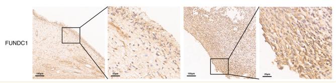
Figure 1 Expressions of inflammatory cytokines, HIF-1α and FUNDC1 in healthy and pulpitis tissues. (A) mRNA expression of IL-1β, IL-6, IL-8 and TNF-α in human healthy and pulpitis tissues. mRNA expression of (B) HIF-1α and (C) FUNDC1 in human healthy and pulpitis tissues. (D) Representative immunostaining images of HIF-1α and FUNDC1 in human healthy or inflamed dental pulp tissues. Scale bars are 100 and 25 µm, respectively. Results are presented as the means ± SD from ≥ three independent experiments. *P<0.05, **P<0.01 and ***P<0.001 vs. healthy. HIF-1α, hypoxia-inducible factor-1α; FUNDC1, FUN14 domain-containing 1.

もっと読みます

Restrictive clause


Affinity Biosciences tests all products strictly. Citations are provided as a resource for additional applications that have not been validated by Affinity Biosciences. Please choose the appropriate format for each application and consult Materials and Methods sections for additional details about the use of any product in these publications.

For Research Use Only.
Not for use in diagnostic or therapeutic procedures. Not for resale. Not for distribution without written consent. Affinity Biosciences will not be held responsible for patent infringement or other violations that may occur with the use of our products. Affinity Biosciences, Affinity Biosciences Logo and all other trademarks are the property of Affinity Biosciences LTD.