製品: SNAP23 Antibody
カタログ: DF13314
タンパク質の説明: Rabbit polyclonal antibody to SNAP23
アプリケーション: WB IHC IF/ICC
反応性: Human, Mouse, Rat
予測: Pig, Bovine, Horse, Sheep, Rabbit, Dog, Chicken
分子量: 23kDa, (isoform)19 kDa; 23kD(Calculated).
ユニプロット: O00161
RRID: AB_2846333

類似製品を見る>>

   サイズ 価格 在庫状況
 100ul $280 在庫あり
 200ul $350 在庫あり

リードタイム: 当日配達

For pricing and ordering contact:
お問い合わせ先

製品説明

ソース:
Rabbit
アプリケーション:
WB 1:500-1:2000, IF/ICC 1:100-1:500, IHC 1:50-1:200
*The optimal dilutions should be determined by the end user.
*Tips:

WB: For western blot detection of denatured protein samples. IHC: For immunohistochemical detection of paraffin sections (IHC-p) or frozen sections (IHC-f) of tissue samples. IF/ICC: For immunofluorescence detection of cell samples. ELISA(peptide): For ELISA detection of antigenic peptide.

反応性:
Human,Mouse,Rat
予測:
Pig(100%), Bovine(100%), Horse(100%), Sheep(100%), Rabbit(100%), Dog(100%), Chicken(83%)
クローナリティ:
Polyclonal
特異性:
SNAP23 Antibody detects endogenous levels of total SNAP23.
RRID:
AB_2846333
引用形式: Affinity Biosciences Cat# DF13314, RRID:AB_2846333.
コンジュゲート:
Unconjugated.
精製:
The antiserum was purified by peptide affinity chromatography using SulfoLink™ Coupling Resin (Thermo Fisher Scientific).
保存:
Rabbit IgG in phosphate buffered saline , pH 7.4, 150mM NaCl, 0.02% sodium azide and 50% glycerol. Store at -20 °C. Stable for 12 months from date of receipt.
別名:

折りたたみ/展開

HsT17016; LS-B8340; SNAP 23; SNAP-23; SNAP23; SNAP23A; SNAP23B; SNP23_HUMAN; Synaptosomal associated protein 23; Synaptosomal associated protein 23kDa; Synaptosomal associated protein; Synaptosomal-associated protein 23; Vesicle membrane fusion protein SNAP 23; Vesicle membrane fusion protein SNAP23; Vesicle-membrane fusion protein SNAP-23;

免疫原

免疫原:
Uniprot:
遺伝子(ID):
発現特異性:
O00161 SNP23_HUMAN:

Ubiquitous. Highest levels where found in placenta.

タンパク質配列:
MDNLSSEEIQQRAHQITDESLESTRRILGLAIESQDAGIKTITMLDEQKEQLNRIEEGLDQINKDMRETEKTLTELNKCCGLCVCPCNRTKNFESGKAYKTTWGDGGENSPCNVVSKQPGPVTNGQLQQPTTGAASGGYIKRITNDAREDEMEENLTQVGSILGNLKDMALNIGNEIDAQNPQIKRITDKADTNRDRIDIANARAKKLIDS

種類予測

種類予測:

Score>80(red) has high confidence and is suggested to be used for WB detection. *The prediction model is mainly based on the alignment of immunogen sequences, the results are for reference only, not as the basis of quality assurance.

Species
Results
Score
Pig
100
Horse
100
Bovine
100
Sheep
100
Dog
100
Rabbit
100
Chicken
83
Xenopus
57
Zebrafish
0
Model Confidence:
High(score>80) Medium(80>score>50) Low(score<50) No confidence

PTMs - O00161 基板として

Site PTM Type Enzyme
M1 Acetylation
S5 Phosphorylation
S6 Phosphorylation
T17 Phosphorylation
S20 Phosphorylation
S23 Phosphorylation P17252 (PRKCA)
T24 Phosphorylation P17252 (PRKCA)
S34 Phosphorylation
K40 Ubiquitination
K49 Ubiquitination
K64 Ubiquitination
K71 Ubiquitination
T72 Phosphorylation
K91 Ubiquitination
S95 Phosphorylation P14618 (PKM)
K97 Ubiquitination
Y99 Phosphorylation
K100 Ubiquitination
T102 Phosphorylation
S110 Phosphorylation
C112 S-Nitrosylation
K117 Ubiquitination
S136 Phosphorylation
Y139 Phosphorylation
K141 Ubiquitination
T144 Phosphorylation
T157 Phosphorylation
S161 Phosphorylation P17252 (PRKCA)
K185 Ubiquitination
K190 Ubiquitination

研究背景

機能:

Essential component of the high affinity receptor for the general membrane fusion machinery and an important regulator of transport vesicle docking and fusion.

細胞の位置付け:

Cell membrane>Peripheral membrane protein. Cell membrane>Lipid-anchor. Cell junction>Synapse>Synaptosome.
Note: Mainly localized to the plasma membrane.

Extracellular region or secreted Cytosol Plasma membrane Cytoskeleton Lysosome Endosome Peroxisome ER Golgi apparatus Nucleus Mitochondrion Manual annotation Automatic computational assertionSubcellular location
組織特異性:

Ubiquitous. Highest levels where found in placenta.

サブユニット構造:

Homotetramer (via coiled-coil domain), also forms heterotetramers with STX4 and VAMP3. Found in a complex with VAMP8 and STX1A. Found in a complex with VAMP8 and STX4 in pancreas (By similarity). Interacts simultaneously with SNAPIN and SYN4 (By similarity). Interacts with STX1A (By similarity). Interacts with STX12 (By similarity). Interacts tightly to multiple syntaxins and synaptobrevins/VAMPs (By similarity). Interacts with ZDHHC13 (via ANK repeats) (By similarity). Interacts with ZDHHC17 (via ANK repeats).

タンパク質ファミリー:

Belongs to the SNAP-25 family.

研究領域

· Genetic Information Processing > Folding, sorting and degradation > SNARE interactions in vesicular transport.

· Organismal Systems > Immune system > Platelet activation.   (View pathway)

参考文献

1). Obstruction of the Tear Drainage Altered Lacrimal Gland Structure and Function. Investigative Ophthalmology & Visual Science (PubMed: 37440262) [IF=4.4]

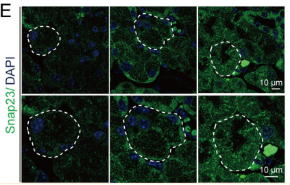
Application: IF/ICC    Species: Mouse    Sample:

Figure 7. Altered expression and localization of proteins relevant to lacrimal gland fusion and secretion. (A) Rab3d immunofluorescence was abundant in the apical lumen side of the acinar cells in the sham LGs (left panels), but uniformly distributed in the STDOB (middle panels) and more basally localized in the Prickle1 mutant acini. The bottom panels are magnified images from the top panels. Apical and basal positions of the acinus lumen are illustrated in the right corner of the first panel. (B) Western blotting demonstrated reduced Rab3d expression in the STDOB and Prickle1 mutant LGs compared with the sham. (C) Vamp8 immunofluorescence showed a similar altered pattern as the Rab3d in the STDOB and Prickle1 mice. (D) Western blotting showed increased LG expression of Vamp8 in the STDOB and Prickle1 mice. (E) Expression of Snap23 in LG acini was also basally shifted and showed increased levels in STDOB and Prickle1 mice compared with the sham (F). The white dotted lines indicate acini boundaries. For Western blotting and quantification, n = 4 LGs/mice.

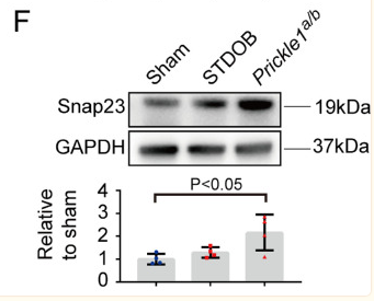
Application: WB    Species: Mouse    Sample:

Figure 7. Altered expression and localization of proteins relevant to lacrimal gland fusion and secretion. (A) Rab3d immunofluorescence was abundant in the apical lumen side of the acinar cells in the sham LGs (left panels), but uniformly distributed in the STDOB (middle panels) and more basally localized in the Prickle1 mutant acini. The bottom panels are magnified images from the top panels. Apical and basal positions of the acinus lumen are illustrated in the right corner of the first panel. (B) Western blotting demonstrated reduced Rab3d expression in the STDOB and Prickle1 mutant LGs compared with the sham. (C) Vamp8 immunofluorescence showed a similar altered pattern as the Rab3d in the STDOB and Prickle1 mice. (D) Western blotting showed increased LG expression of Vamp8 in the STDOB and Prickle1 mice. (E) Expression of Snap23 in LG acini was also basally shifted and showed increased levels in STDOB and Prickle1 mice compared with the sham (F). The white dotted lines indicate acini boundaries. For Western blotting and quantification, n = 4 LGs/mice.

Restrictive clause


Affinity Biosciences tests all products strictly. Citations are provided as a resource for additional applications that have not been validated by Affinity Biosciences. Please choose the appropriate format for each application and consult Materials and Methods sections for additional details about the use of any product in these publications.

For Research Use Only.
Not for use in diagnostic or therapeutic procedures. Not for resale. Not for distribution without written consent. Affinity Biosciences will not be held responsible for patent infringement or other violations that may occur with the use of our products. Affinity Biosciences, Affinity Biosciences Logo and all other trademarks are the property of Affinity Biosciences LTD.