製品: RAB12 Antibody
カタログ: DF12459
タンパク質の説明: Rabbit polyclonal antibody to RAB12
アプリケーション: WB IHC
Cited expt.: WB
反応性: Human, Mouse
予測: Pig, Bovine, Horse, Sheep, Rabbit, Dog, Chicken
分子量: 28 kDa; 27kD(Calculated).
ユニプロット: Q6IQ22
RRID: AB_2845264

類似製品を見る>>

   サイズ 価格 在庫状況
 100ul $280 在庫あり
 200ul $350 在庫あり

リードタイム: 当日配達

For pricing and ordering contact:
お問い合わせ先

製品説明

ソース:
Rabbit
アプリケーション:
WB 1:500-1:2000, IHC 1:50-1:200
*The optimal dilutions should be determined by the end user. For optimal experimental results, antibody reuse is not recommended.
*Tips:

WB: For western blot detection of denatured protein samples. IHC: For immunohistochemical detection of paraffin sections (IHC-p) or frozen sections (IHC-f) of tissue samples. IF/ICC: For immunofluorescence detection of cell samples. ELISA(peptide): For ELISA detection of antigenic peptide.

反応性:
Human,Mouse
予測:
Pig(100%), Bovine(100%), Horse(100%), Sheep(100%), Rabbit(100%), Dog(100%), Chicken(100%)
クローナリティ:
Polyclonal
特異性:
RAB12 Antibody detects endogenous levels of total RAB12.
RRID:
AB_2845264
引用形式: Affinity Biosciences Cat# DF12459, RRID:AB_2845264.
コンジュゲート:
Unconjugated.
精製:
The antiserum was purified by peptide affinity chromatography using SulfoLink™ Coupling Resin (Thermo Fisher Scientific).
保存:
Rabbit IgG in phosphate buffered saline , pH 7.4, 150mM NaCl, 0.02% sodium azide and 50% glycerol. Store at -20 °C. Stable for 12 months from date of receipt.
別名:

折りたたみ/展開

Putative Ras related protein Rab 12; RAB12; RAB12, member RAS oncogene family; RAB12_HUMAN; Ras-related protein Rab-12;

免疫原

免疫原:

A synthesized peptide derived from human RAB12, corresponding to a region within C-terminal amino acids.

Uniprot:
遺伝子(ID):
タンパク質配列:
MDPGAALQRRAGGGGGLGAGSPALSGGQGRRRKQPPRPADFKLQVIIIGSRGVGKTSLMERFTDDTFCEACKSTVGVDFKIKTVELRGKKIRLQIWDTAGQERFNSITSAYYRSAKGIILVYDITKKETFDDLPKWMKMIDKYASEDAELLLVGNKLDCETDREITRQQGEKFAQQITGMRFCEASAKDNFNVDEIFLKLVDDILKKMPLDILRNELSNSILSLQPEPEIPPELPPPRPHVRCC

種類予測

種類予測:

Score>80(red) has high confidence and is suggested to be used for WB detection. *The prediction model is mainly based on the alignment of immunogen sequences, the results are for reference only, not as the basis of quality assurance.

Species
Results
Score
Pig
100
Horse
100
Bovine
100
Sheep
100
Dog
100
Chicken
100
Rabbit
100
Xenopus
0
Zebrafish
0
Model Confidence:
High(score>80) Medium(80>score>50) Low(score<50) No confidence

研究背景

機能:

The small GTPases Rab are key regulators of intracellular membrane trafficking, from the formation of transport vesicles to their fusion with membranes. Rabs cycle between an inactive GDP-bound form and an active GTP-bound form that is able to recruit to membranes different set of downstream effectors directly responsible for vesicle formation, movement, tethering and fusion. That Rab may play a role in protein transport from recycling endosomes to lysosomes regulating, for instance, the degradation of the transferrin receptor. Involved in autophagy (By similarity).

PTMs:

Phosphorylation of Ser-106 in the switch II region by LRRK2 prevents the association of RAB regulatory proteins, including CHM, CHML and RAB GDP dissociation inhibitors GDI1 and GDI2.

細胞の位置付け:

Recycling endosome membrane>Lipid-anchor>Cytoplasmic side. Lysosome membrane>Lipid-anchor>Cytoplasmic side. Golgi apparatus membrane. Cytoplasmic vesicle>Autophagosome.

Extracellular region or secreted Cytosol Plasma membrane Cytoskeleton Lysosome Endosome Peroxisome ER Golgi apparatus Nucleus Mitochondrion Manual annotation Automatic computational assertionSubcellular location
タンパク質ファミリー:

Belongs to the small GTPase superfamily. Rab family.

参考文献

1). TMT-based quantitative proteomics analysis reveals the attenuated replication mechanism of Newcastle disease virus caused by nuclear localization signal mutation in viral matrix protein. Virulence, 2020 (PubMed: 32420802) [IF=5.5]

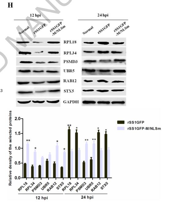
Application: WB    Species: Mouse    Sample: BSR-T7/5 cells

Figure 8. rSS1GFP infection affects the expression of cellular translation, posttranslational modification and trafficking-associated proteins. (A) The heatmap of representative 20 DEPs related to “Translation, ribosomal structure and biogenesis” during rSS1GFP and rSS1GFP-M/NLSm infection at 12 and 24 h. (B) The protein-protein interactions of the DEPs related to “Translation, ribosomal structure and biogenesis” are analyzed by the STRING software. A red line indicates the presence of fusion evidence; a blue line indicates co-occurrence evidence; a light blue line indicates database evidence; a purple line indicates experimental evidence; a green line indicates neighborhood evidence; a black line indicates co-expression evidence. (C) The heatmap of representative 20 DEPs related to “Posttranslational modification, protein turnover, chaperones” during rSS1GFP and rSS1GFP-M/NLSm infection at 12 and 24 h. (D) The protein-protein interactions of the DEPs related to “Posttranslational modification, protein turnover, chaperones” are analyzed by the STRING software. (E) The heatmap of representative 20 DEPs related to “Intracellular trafficking, secretion, and vesicular transport” during rSS1GFP and rSS1GFP-M/NLSm infection at 12 and 24 h. (F) The protein-protein interactions of the DEPs related to “Intracellular trafficking, secretion, and vesicular transport” are analyzed by the STRING software. (G) The mRNA expression levels of six selected DEP genes in BSR-T7/5 cells infected with rSS1GFP and rSS1GFP-M/NLSm were verified by qRT-PCR. (H) The protein expression levels of six DEPs in BSR-T7/5 cells infected with rSS1GFP and rSS1GFP-M/NLSm were examined by Western blotting. The relative expression levels of six DEPs were compared with the control GAPDH expression.

Restrictive clause


Affinity Biosciences tests all products strictly. Citations are provided as a resource for additional applications that have not been validated by Affinity Biosciences. Please choose the appropriate format for each application and consult Materials and Methods sections for additional details about the use of any product in these publications.

For Research Use Only.
Not for use in diagnostic or therapeutic procedures. Not for resale. Not for distribution without written consent. Affinity Biosciences will not be held responsible for patent infringement or other violations that may occur with the use of our products. Affinity Biosciences, Affinity Biosciences Logo and all other trademarks are the property of Affinity Biosciences LTD.