製品: SYVN1 Antibody
カタログ: DF12235
タンパク質の説明: Rabbit polyclonal antibody to SYVN1
アプリケーション: WB IHC IF/ICC
反応性: Human, Mouse, Rat
予測: Pig, Bovine, Horse, Sheep, Rabbit, Dog
分子量: 68-76 kDa; 68kD(Calculated).
ユニプロット: Q86TM6
RRID: AB_2845040

類似製品を見る>>

   サイズ 価格 在庫状況
 100ul $280 在庫あり
 200ul $350 在庫あり

リードタイム: 当日配達

For pricing and ordering contact:
お問い合わせ先

製品説明

ソース:
Rabbit
アプリケーション:
WB 1:500-1:2000, IHC 1:50-1:200, IF/ICC 1:100-1:500
*The optimal dilutions should be determined by the end user.
*Tips:

WB: For western blot detection of denatured protein samples. IHC: For immunohistochemical detection of paraffin sections (IHC-p) or frozen sections (IHC-f) of tissue samples. IF/ICC: For immunofluorescence detection of cell samples. ELISA(peptide): For ELISA detection of antigenic peptide.

反応性:
Human,Mouse,Rat
予測:
Pig(100%), Bovine(100%), Horse(100%), Sheep(100%), Rabbit(100%), Dog(100%)
クローナリティ:
Polyclonal
特異性:
SYVN1 Antibody detects endogenous levels of total SYVN1.
RRID:
AB_2845040
引用形式: Affinity Biosciences Cat# DF12235, RRID:AB_2845040.
コンジュゲート:
Unconjugated.
精製:
The antiserum was purified by peptide affinity chromatography using SulfoLink™ Coupling Resin (Thermo Fisher Scientific).
保存:
Rabbit IgG in phosphate buffered saline , pH 7.4, 150mM NaCl, 0.02% sodium azide and 50% glycerol. Store at -20 °C. Stable for 12 months from date of receipt.
別名:

折りたたみ/展開

1200010C09Rik; DER3; E3 ubiquitin-protein ligase synoviolin; HMG coA reductase degradation 1 homolog; HRD1; KIAA1810; MGC40372; OTTHUMP00000230429; OTTHUMP00000230430; OTTHUMP00000230431; OTTHUMP00000230432; Synovial apoptosis inhibitor 1; Synovial apoptosis inhibitor 1, synoviolin; Synoviolin 1 isoform b; SYNOVIOLIN; SYVN1; SYVN1_HUMAN;

免疫原

免疫原:
Uniprot:
遺伝子(ID):
発現特異性:
Q86TM6 SYVN1_HUMAN:

Ubiquitously expressed, with highest levels in liver and kidney (at protein level). Up-regulated in synovial tissues from patients with rheumatoid arthritis (at protein level).

タンパク質配列:
MFRTAVMMAASLALTGAVVAHAYYLKHQFYPTVVYLTKSSPSMAVLYIQAFVLVFLLGKVMGKVFFGQLRAAEMEHLLERSWYAVTETCLAFTVFRDDFSPRFVALFTLLLFLKCFHWLAEDRVDFMERSPNISWLFHCRIVSLMFLLGILDFLFVSHAYHSILTRGASVQLVFGFEYAILMTMVLTIFIKYVLHSVDLQSENPWDNKAVYMLYTELFTGFIKVLLYMAFMTIMIKVHTFPLFAIRPMYLAMRQFKKAVTDAIMSRRAIRNMNTLYPDATPEELQAMDNVCIICREEMVTGAKRLPCNHIFHTSCLRSWFQRQQTCPTCRMDVLRASLPAQSPPPPEPADQGPPPAPHPPPLLPQPPNFPQGLLPPFPPGMFPLWPPMGPFPPVPPPPSSGEAVAPPSTSAAALSRPSGAATTTAAGTSATAASATASGPGSGSAPEAGPAPGFPFPPPWMGMPLPPPFAFPPMPVPPAGFAGLTPEELRALEGHERQHLEARLQSLRNIHTLLDAAMLQINQYLTVLASLGPPRPATSVNSTEETATTVVAAASSTSIPSSEATTPTPGASPPAPEMERPPAPESVGTEEMPEDGEPDAAELRRRRLQKLESPVAH

種類予測

種類予測:

Score>80(red) has high confidence and is suggested to be used for WB detection. *The prediction model is mainly based on the alignment of immunogen sequences, the results are for reference only, not as the basis of quality assurance.

Species
Results
Score
Pig
100
Horse
100
Bovine
100
Sheep
100
Dog
100
Rabbit
100
Xenopus
67
Zebrafish
67
Chicken
0
Model Confidence:
High(score>80) Medium(80>score>50) Low(score<50) No confidence

PTMs - Q86TM6 基板として

Site PTM Type Enzyme
T93 Phosphorylation
K191 Ubiquitination
K257 Ubiquitination
T260 Phosphorylation
S265 Phosphorylation
K303 Ubiquitination
T557 Phosphorylation
S558 Phosphorylation
S562 Phosphorylation
T565 Phosphorylation
S572 Phosphorylation
S613 Phosphorylation

研究背景

機能:

Acts as an E3 ubiquitin-protein ligase which accepts ubiquitin specifically from endoplasmic reticulum-associated UBC7 E2 ligase and transfers it to substrates, promoting their degradation. Component of the endoplasmic reticulum quality control (ERQC) system also called ER-associated degradation (ERAD) involved in ubiquitin-dependent degradation of misfolded endoplasmic reticulum proteins. Also promotes the degradation of normal but naturally short-lived proteins such as SGK. Protects cells from ER stress-induced apoptosis. Protects neurons from apoptosis induced by polyglutamine-expanded huntingtin (HTT) or unfolded GPR37 by promoting their degradation. Sequesters p53/TP53 in the cytoplasm and promotes its degradation, thereby negatively regulating its biological function in transcription, cell cycle regulation and apoptosis. Mediates the ubiquitination and subsequent degradation of cytoplasmic NFE2L1 (By similarity).

PTMs:

Not N-glycosylated.

Auto-ubiquitinated.

細胞の位置付け:

Endoplasmic reticulum membrane>Multi-pass membrane protein.

Extracellular region or secreted Cytosol Plasma membrane Cytoskeleton Lysosome Endosome Peroxisome ER Golgi apparatus Nucleus Mitochondrion Manual annotation Automatic computational assertionSubcellular location
組織特異性:

Ubiquitously expressed, with highest levels in liver and kidney (at protein level). Up-regulated in synovial tissues from patients with rheumatoid arthritis (at protein level).

サブユニット構造:

Homodimer. Interacts with p53/TP53 and HTT. Interacts with VCP, HERPUD1 and DERL1. Part of a complex containing SYVN1, HERPUD1, VIMP and DERL1; which probably transfer misfolded proteins from the ER to VCP. Part of a complex containing SYVN1, SEL1L and DERL2. Interacts with UBXN6. Interacts (via N-terminal loop) with SEL1L; recruits ERLEC1 and OS9. May form a complex with ERLEC1; HSPA5; OS9 AND SEL1L. Interacts with BAG6. Interacts with NFE2L1 (By similarity).

タンパク質ファミリー:

The RING-type zinc finger is required for E3 ligase activity.

Belongs to the HRD1 family.

研究領域

· Genetic Information Processing > Folding, sorting and degradation > Ubiquitin mediated proteolysis.   (View pathway)

· Genetic Information Processing > Folding, sorting and degradation > Protein processing in endoplasmic reticulum.   (View pathway)

参考文献

1). Endoplasmic reticulum stress-triggered ferroptosis via the XBP1-Hrd1-Nrf2 pathway induces EMT progression in diabetic nephropathy.. Biomedicine & Pharmacotherapy (PubMed: 37224754) [IF=7.5]

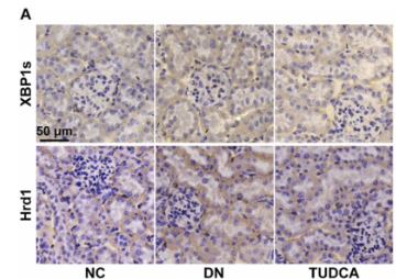
Application: IHC    Species: Mouse    Sample: kidney tissues

Fig. 6. The XBP1-Hrd1 arm of the ERS pathway was triggered by high glucose. The XBP1s and Hrd1 expression in tissue samples was detected by immunohistochemical staining (n = 3) (A). Semi-quantitative analysis of XBP1s and Hrd1 expression in each group (n = 3) (B). The qRT-PCR results of Hrd1 in NC, DM, TUDCA, and PBS group (n = 3) (C). The protein expression of Hrd1 in each group of mice (n = 3) (D - E). The fluorescence intensity of XBP1s and Hrd1 in HK-2 cells was measured by the confocal fluorescence microscope. Blue represented the cell nucleus and red represented XBP1s or Hrd1 (n = 3) (F - G). Fluorescence intensity was quantified using Image J (n = 3) (H - I). The mRNA level of Hrd1 among Ctrl, HG, Mannitol, TUDCA, and DMSO groups (n = 3) (J). Immunoblot and semi-quantitative analysis of Hrd1 in each group cells (n = 3) (K - L). The qRT-PCR results of XBP1s and Hrd1 in cells transfected with empty vectors or XBP1 overexpression plasmids. At 6 h post-transfection, cells were changed to a medium containing 30 mmol/L glucose with or without TUDCA (200 μM) for 48 h (n = 3) (M). Immunoblot and quantitative analysis of XBP1s and Hrd1 under the same conditions (n = 3) (N - O). Data are expressed as the mean ± SEM.

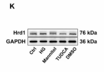
Application: WB    Species: Human    Sample: HK-2 cells

Fig. 6. The XBP1-Hrd1 arm of the ERS pathway was triggered by high glucose. The XBP1s and Hrd1 expression in tissue samples was detected by immunohistochemical staining (n = 3) (A). Semi-quantitative analysis of XBP1s and Hrd1 expression in each group (n = 3) (B). The qRT-PCR results of Hrd1 in NC, DM, TUDCA, and PBS group (n = 3) (C). The protein expression of Hrd1 in each group of mice (n = 3) (D - E). The fluorescence intensity of XBP1s and Hrd1 in HK-2 cells was measured by the confocal fluorescence microscope. Blue represented the cell nucleus and red represented XBP1s or Hrd1 (n = 3) (F - G). Fluorescence intensity was quantified using Image J (n = 3) (H - I). The mRNA level of Hrd1 among Ctrl, HG, Mannitol, TUDCA, and DMSO groups (n = 3) (J). Immunoblot and semi-quantitative analysis of Hrd1 in each group cells (n = 3) (K - L). The qRT-PCR results of XBP1s and Hrd1 in cells transfected with empty vectors or XBP1 overexpression plasmids. At 6 h post-transfection, cells were changed to a medium containing 30 mmol/L glucose with or without TUDCA (200 μM) for 48 h (n = 3) (M). Immunoblot and quantitative analysis of XBP1s and Hrd1 under the same conditions (n = 3) (N - O). Data are expressed as the mean ± SEM.

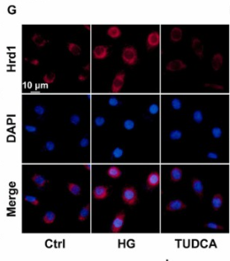
Application: IF/ICC    Species: Human    Sample: HK-2 cells

Fig. 6. The XBP1-Hrd1 arm of the ERS pathway was triggered by high glucose. The XBP1s and Hrd1 expression in tissue samples was detected by immunohistochemical staining (n = 3) (A). Semi-quantitative analysis of XBP1s and Hrd1 expression in each group (n = 3) (B). The qRT-PCR results of Hrd1 in NC, DM, TUDCA, and PBS group (n = 3) (C). The protein expression of Hrd1 in each group of mice (n = 3) (D - E). The fluorescence intensity of XBP1s and Hrd1 in HK-2 cells was measured by the confocal fluorescence microscope. Blue represented the cell nucleus and red represented XBP1s or Hrd1 (n = 3) (F - G). Fluorescence intensity was quantified using Image J (n = 3) (H - I). The mRNA level of Hrd1 among Ctrl, HG, Mannitol, TUDCA, and DMSO groups (n = 3) (J). Immunoblot and semi-quantitative analysis of Hrd1 in each group cells (n = 3) (K - L). The qRT-PCR results of XBP1s and Hrd1 in cells transfected with empty vectors or XBP1 overexpression plasmids. At 6 h post-transfection, cells were changed to a medium containing 30 mmol/L glucose with or without TUDCA (200 μM) for 48 h (n = 3) (M). Immunoblot and quantitative analysis of XBP1s and Hrd1 under the same conditions (n = 3) (N - O). Data are expressed as the mean ± SEM.

Restrictive clause


Affinity Biosciences tests all products strictly. Citations are provided as a resource for additional applications that have not been validated by Affinity Biosciences. Please choose the appropriate format for each application and consult Materials and Methods sections for additional details about the use of any product in these publications.

For Research Use Only.
Not for use in diagnostic or therapeutic procedures. Not for resale. Not for distribution without written consent. Affinity Biosciences will not be held responsible for patent infringement or other violations that may occur with the use of our products. Affinity Biosciences, Affinity Biosciences Logo and all other trademarks are the property of Affinity Biosciences LTD.