Phospho-STAT5 (Ser725/Ser730)[Ser726/Ser731] Antibody - #AF3304
 (8)  
			
			
			(12)
(8)  
			
			
			(12)  
			
			
		| 製品: | Phospho-STAT5 (Ser725/Ser730)[Ser726/Ser731] Antibody | 
| カタログ: | AF3304 | 
| タンパク質の説明: | Rabbit polyclonal antibody to Phospho-STAT5 (Ser725/Ser730)[Ser726/Ser731] | 
| アプリケーション: | WB IHC IF/ICC | 
| Cited expt.: | WB, IHC, IF/ICC | 
| 反応性: | Human, Mouse, Rat | 
| 分子量: | 90kDa; 91kD,90kD(Calculated). | 
| ユニプロット: | P42229 | P51692 | 
| RRID: | AB_2834723 | 
製品説明
*The optimal dilutions should be determined by the end user.
*Tips:
WB: For western blot detection of denatured protein samples. IHC: For immunohistochemical detection of paraffin sections (IHC-p) or frozen sections (IHC-f) of tissue samples. IF/ICC: For immunofluorescence detection of cell samples. ELISA(peptide): For ELISA detection of antigenic peptide.
引用形式: Affinity Biosciences Cat# AF3304, RRID:AB_2834723.
折りたたみ/展開
Mammary gland factor; MGF; Signal transducer and activator of transcription 5A; STA5A_HUMAN; STAT 5; STAT 5A; STAT5; STAT5A; Signal transducer and activator of transcription 5B; STA5B_HUMAN; STAT5; Stat5b; Transcription factor STAT5B;
免疫原
A synthesized peptide derived from human STAT5 around the phosphorylation site of Ser725/Ser730.
- P42229 STA5A_HUMAN:
- Protein BLAST With
- NCBI/
- ExPASy/
- Uniprot
MAGWIQAQQLQGDALRQMQVLYGQHFPIEVRHYLAQWIESQPWDAIDLDNPQDRAQATQLLEGLVQELQKKAEHQVGEDGFLLKIKLGHYATQLQKTYDRCPLELVRCIRHILYNEQRLVREANNCSSPAGILVDAMSQKHLQINQTFEELRLVTQDTENELKKLQQTQEYFIIQYQESLRIQAQFAQLAQLSPQERLSRETALQQKQVSLEAWLQREAQTLQQYRVELAEKHQKTLQLLRKQQTIILDDELIQWKRRQQLAGNGGPPEGSLDVLQSWCEKLAEIIWQNRQQIRRAEHLCQQLPIPGPVEEMLAEVNATITDIISALVTSTFIIEKQPPQVLKTQTKFAATVRLLVGGKLNVHMNPPQVKATIISEQQAKSLLKNENTRNECSGEILNNCCVMEYHQATGTLSAHFRNMSLKRIKRADRRGAESVTEEKFTVLFESQFSVGSNELVFQVKTLSLPVVVIVHGSQDHNATATVLWDNAFAEPGRVPFAVPDKVLWPQLCEALNMKFKAEVQSNRGLTKENLVFLAQKLFNNSSSHLEDYSGLSVSWSQFNRENLPGWNYTFWQWFDGVMEVLKKHHKPHWNDGAILGFVNKQQAHDLLINKPDGTFLLRFSDSEIGGITIAWKFDSPERNLWNLKPFTTRDFSIRSLADRLGDLSYLIYVFPDRPKDEVFSKYYTPVLAKAVDGYVKPQIKQVVPEFVNASADAGGSSATYMDQAPSPAVCPQAPYNMYPQNPDHVLDQDGEFDLDETMDVARHVEELLRRPMDSLDSRLSPPAGLFTSARGSLS
- P51692 STA5B_HUMAN:
- Protein BLAST With
- NCBI/
- ExPASy/
- Uniprot
MAVWIQAQQLQGEALHQMQALYGQHFPIEVRHYLSQWIESQAWDSVDLDNPQENIKATQLLEGLVQELQKKAEHQVGEDGFLLKIKLGHYATQLQNTYDRCPMELVRCIRHILYNEQRLVREANNGSSPAGSLADAMSQKHLQINQTFEELRLVTQDTENELKKLQQTQEYFIIQYQESLRIQAQFGPLAQLSPQERLSRETALQQKQVSLEAWLQREAQTLQQYRVELAEKHQKTLQLLRKQQTIILDDELIQWKRRQQLAGNGGPPEGSLDVLQSWCEKLAEIIWQNRQQIRRAEHLCQQLPIPGPVEEMLAEVNATITDIISALVTSTFIIEKQPPQVLKTQTKFAATVRLLVGGKLNVHMNPPQVKATIISEQQAKSLLKNENTRNDYSGEILNNCCVMEYHQATGTLSAHFRNMSLKRIKRSDRRGAESVTEEKFTILFESQFSVGGNELVFQVKTLSLPVVVIVHGSQDNNATATVLWDNAFAEPGRVPFAVPDKVLWPQLCEALNMKFKAEVQSNRGLTKENLVFLAQKLFNNSSSHLEDYSGLSVSWSQFNRENLPGRNYTFWQWFDGVMEVLKKHLKPHWNDGAILGFVNKQQAHDLLINKPDGTFLLRFSDSEIGGITIAWKFDSQERMFWNLMPFTTRDFSIRSLADRLGDLNYLIYVFPDRPKDEVYSKYYTPVPCESATAKAVDGYVKPQIKQVVPEFVNASADAGGGSATYMDQAPSPAVCPQAHYNMYPQNPDSVLDTDGDFDLEDTMDVARRVEELLGRPMDSQWIPHAQS
研究背景
Carries out a dual function: signal transduction and activation of transcription. Mediates cellular responses to the cytokine KITLG/SCF and other growth factors. Mediates cellular responses to ERBB4. May mediate cellular responses to activated FGFR1, FGFR2, FGFR3 and FGFR4. Binds to the GAS element and activates PRL-induced transcription. Regulates the expression of milk proteins during lactation.
Tyrosine phosphorylated in response to KITLG/SCF, IL2, IL3, IL7, IL15, CSF2/GMCSF, GH1, PRL, EPO and THPO (By similarity). Activated KIT promotes phosphorylation on tyrosine residues and subsequent translocation to the nucleus. Tyrosine phosphorylated in response to constitutively activated FGFR1, FGFR2, FGFR3 and FGFR4 (By similarity). Tyrosine phosphorylation is required for DNA-binding activity and dimerization. Serine phosphorylation is also required for maximal transcriptional activity (By similarity). Tyrosine phosphorylated in response to signaling via activated FLT3; wild-type FLT3 results in much weaker phosphorylation than constitutively activated mutant FLT3. Alternatively, can be phosphorylated by JAK2 at Tyr-694.
ISGylated.
Cytoplasm. Nucleus. 
Note: Translocated into the nucleus in response to phosphorylation.
Belongs to the transcription factor STAT family.
Carries out a dual function: signal transduction and activation of transcription. Mediates cellular responses to the cytokine KITLG/SCF and other growth factors. Binds to the GAS element and activates PRL-induced transcription. Positively regulates hematopoietic/erythroid differentiation.
Tyrosine phosphorylated in response to signaling via activated KIT, resulting in translocation to the nucleus. Tyrosine phosphorylated in response to signaling via activated FLT3; wild-type FLT3 results in much weaker phosphorylation than constitutively activated mutant FLT3. Alternatively, can be phosphorylated by JAK2. Phosphorylation at Tyr-699 by PTK6 or HCK leads to an increase of its transcriptional activity.
Cytoplasm. Nucleus. 
Note: Translocated into the nucleus in response to phosphorylation.
Belongs to the transcription factor STAT family.
研究領域
· Cellular Processes > Cell growth and death > Necroptosis. (View pathway)
· Environmental Information Processing > Signal transduction > ErbB signaling pathway. (View pathway)
· Environmental Information Processing > Signal transduction > Jak-STAT signaling pathway. (View pathway)
· Human Diseases > Infectious diseases: Viral > Hepatitis B.
· Human Diseases > Infectious diseases: Viral > Measles.
· Human Diseases > Infectious diseases: Viral > HTLV-I infection.
· Human Diseases > Cancers: Overview > Pathways in cancer. (View pathway)
· Human Diseases > Cancers: Overview > Viral carcinogenesis.
· Human Diseases > Cancers: Specific types > Chronic myeloid leukemia. (View pathway)
· Human Diseases > Cancers: Specific types > Acute myeloid leukemia. (View pathway)
· Human Diseases > Cancers: Specific types > Non-small cell lung cancer. (View pathway)
· Organismal Systems > Immune system > Chemokine signaling pathway. (View pathway)
· Organismal Systems > Immune system > Th1 and Th2 cell differentiation. (View pathway)
· Organismal Systems > Immune system > Th17 cell differentiation. (View pathway)
· Organismal Systems > Endocrine system > Prolactin signaling pathway. (View pathway)
参考文献
Application: IHC Species: rat Sample: kidney
Application: WB Species: rat Sample: kidney
Application: WB Species: Mouse Sample:
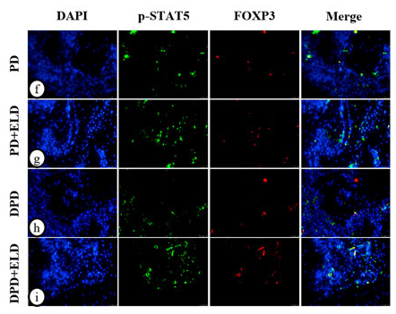
Application: IF/ICC Species: Mouse Sample:
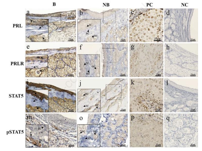
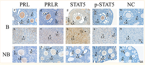
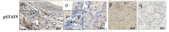
Application: IHC Species: rat Sample: glandular cells, epithelial cells and interstitial cells
Application: IHC Species: Mouse Sample:
Application: IHC Species: muskrats Sample:
Restrictive clause
Affinity Biosciences tests all products strictly. Citations are provided as a resource for additional applications that have not been validated by Affinity Biosciences. Please choose the appropriate format for each application and consult Materials and Methods sections for additional details about the use of any product in these publications.
For Research Use Only.
Not for use in diagnostic or therapeutic procedures. Not for resale. Not for distribution without written consent. Affinity Biosciences will not be held responsible for patent infringement or other violations that may occur with the use of our products. Affinity Biosciences, Affinity Biosciences Logo and all other trademarks are the property of Affinity Biosciences LTD.
![AF3304 at 1/100 staining human breast carcinoma tissue by IHC-P. The sample was formaldehyde fixed and a heat mediated antigen retrieval step in citrate buffer was performed. The sample was then blocked and incubated with the antibody for 1.5 hours at 22°C. An HRP conjugated goat anti-rabbit antibody was used as the secondary antibody. Phospho-STAT5 (Ser725/Ser730)[Ser726/Ser731] Antibody - AF3304 at 1/100 staining human breast carcinoma tissue by IHC-P.](http://img.affbiotech.cn/images/201708/thumb_img/1469_thumb_P_1502255682426.jpg) 
											![AF3304 staining HeLa cells by ICC/IF. Cells were fixed with PFA and permeabilized in 0.1% saponin prior to blocking in 10% serum for 45 minutes at 37°C. The primary antibody was diluted 1/400 and incubated with the sample for 1 hour at 37°C. A  Alexa Fluor 594 conjugated goat polyclonal to rabbit IgG (H+L), diluted 1/600 was used as secondary antibody. Phospho-STAT5 (Ser725/Ser730)[Ser726/Ser731] Antibody - AF3304 staining HeLa cells by ICC/IF.](http://img.affbiotech.cn/images/201708/thumb_img/1469_thumb_P_1503454258006.jpg) 
											![AF3304 staining Hela cells(4h of LPS treatment) by IF/ICC. The samples were fixed with PFA and permeabilized in 0.1% Triton X-100,then blocked in 10% serum for 45 minutes at 25°C. Samples were then incubated with primary Ab(AF3304 1:200) and mouse anti-beta tubulin Ab(T0023 1:200) for 1 hour at 37°C. An  AlexaFluor594 conjugated goat anti-rabbit IgG(H+L) Ab(Red) and an AlexaFluor488 conjugated goat anti-mouse IgG(H+L) Ab(Green) were used as the secondary antibody.
The nuclear counter stain is DAPI(blue). Phospho-STAT5 (Ser725/Ser730)[Ser726/Ser731] Antibody - AF3304 staining Hela cells(4h of LPS treatment) by IF/ICC.](http://img.affbiotech.cn/images/201907/thumb_img/af3304_phospho_stat5ab_ser725730_antibody_thumb_P_1562429765014.jpg) 
											![peptide-ELISA analysis of AF3304. showing specificity to antigen peptide. Peptides concentration: 1ug/ml.<br>
                    P-peptide: phospho-peptide. N-peptide: non-phospho-peptide.	 Phospho-STAT5 (Ser725/Ser730)[Ser726/Ser731] Antibody - peptide-ELISA analysis of AF3304.](http://img.affbiotech.cn/images/pelisa/809/af3304-peptide-elisa.png) 
											![Figure 3. | Seasonal immunolocalization of PRL, PRLR, STAT5 and pSTAT5 in the scented glands of the muskrats. Black arrows indicatethe three cell types: glandular cells, epithelial cells and interstitial cells. The first column (a,e,i,m) represents staining in the breedingseason. The second column (b,f,j,o) represent staining in the non-breeding season. The third (c, g, k, p) and fourth (d, h, l, q) columnsrepresent staining in the positive and negative control, respectively. a,b) Immunolocalization of PRL in the scented glands. e,f)Immunolocalization of PRLR in the scented glands. i,j) Immunolocalization of STAT5 in the scented glands. m,o) Immunolocalizationof pSTAT5 in the scented glands. PRL, PRLR, STAT5 and pSTAT5 detected in the mammary glands were used as a positive control(c,g,k,p). Negative control sections were treated with normal rabbit serum instead of primary antisera (d,h,l,q). B, breeding season; NB,non-breeding season; PC, positive control; NC, negative control; GC, glandular cell; EC, epithelial cell; IC, interstitial cell. Scale bars:25 μm. Phospho-STAT5 (Ser725/Ser730)[Ser726/Ser731] Antibody - Figure 3.](http://img.affbiotech.cn/images/cited_image/202201/cite-wx-446-1641550585.jpg) 
											![Fig. 7.| Effect of the YGY on the HIF1α-STAT5 pathway in the kidney of CKD rats with reproductive dysfunction. (A–D) Expression of EPO (A), p-EPOR (Tyr368) (B),p-JAK2 (Tyr570) (C), and p-STAT5 (Ser725) (D) were determined via immunohistochemistry. The presence of these proteins is indicated by brown staining as observed via light microscopy. (A1, B1, C1, D1) kidneys of the NC group rats; (A2, B2, C2, D2) kidneys of the CKD group rats; (A3, B3, C3, D3) kidneys of the GL 10 g kg−1 group rats; (A4, B4, C4, D4) kidneys of the YGY 10 g kg−1 group rats; (A5, B5, C5, D5)kidneys of the YGY 20 g kg−1 group rats; (A6, B6, C6, D6) kidneys of the YGY40 g•kg−1 group rats (Bar = 50 μm, 200 × , the red arrow refers to the brown staining). Quantitative data are shown in the right panel. Phospho-STAT5 (Ser725/Ser730)[Ser726/Ser731] Antibody - Fig.](http://img.affbiotech.cn/images/cited_image/202202/cite-wx-881-1644569031.jpg) 
											![Fig. 7.| Effect of the YGY on the HIF1α-STAT5 pathway in the kidney of CKD rats with reproductive dysfunction. (E) Western blotting bands for kidney HIF1α, EPO, p-EPOR (Tyr368), total EPOR, p-JAK2 (Tyr570), total JAK2, p-STAT5 (Ser725) and total STAT5 proteins, and quantitative data are shown in the lower panel. Data are presented as the mean ± SD. (n=3). *P < 0.05, **P < 0.01 compared with the NC group. #P < 0.05, ##P < 0.01 compared with the CKD group. (For interpretation of the references to color in this figure legend, the reader is referred to the Web version of this article.) Phospho-STAT5 (Ser725/Ser730)[Ser726/Ser731] Antibody - Fig.](http://img.affbiotech.cn/images/cited_image/202202/cite-wx-882-1644569031.jpg) 
											![Figure 3. Seasonal immunolocalization of PRL, PRLR, STAT5 and pSTAT5 in the scented glands of the muskrats. Black arrows indicate the three cell types: glandular cells, epithelial cells and interstitial cells. The first column (a,e,i,m) represents staining in the breeding season. The second column (b,f,j,o) represent staining in the non-breeding season. The third (c, g, k, p) and fourth (d, h, l, q) columns represent staining in the positive and negative control, respectively. a,b) Immunolocalization of PRL in the scented glands. e,f) Immunolocalization of PRLR in the scented glands. i,j) Immunolocalization of STAT5 in the scented glands. m,o) Immunolocalization of pSTAT5 in the scented glands. PRL, PRLR, STAT5 and pSTAT5 detected in the mammary glands were used as a positive control (c,g,k,p). Negative control sections were treated with normal rabbit serum instead of primary antisera (d,h,l,q). B, breeding season; NB, non-breeding season; PC, positive control; NC, negative control; GC, glandular cell; EC, epithelial cell; IC, interstitial cell. Scale bars: 25 μm. Phospho-STAT5 (Ser725/Ser730)[Ser726/Ser731] Antibody - Figure 3.](http://img.affbiotech.cn/uploads/202312/bc069f214a65c3292f1e70b4fc44e973.png) 
											![FIGURE 7. ELD regulated Th17/Treg cells through the STAT3/STAT5 signal. (A) (a) The protein expression of p-STAT3, STAT3, p-STAT5, STAT5 and GAPDH detected by western blotting in the PD, PD + ELD, DPD and DPD + ELD groups. The statistical analysis of (b) p-STAT3/STAT3, (c) p-STAT5/STAT5 and (d) the ratio of p-STAT3/p-STAT5 in western blotting. (B) The representative immunofluorescence staining images of p-STAT3+IL-17A+ cells in the (a) PD, (b) PD + ELD, (c) DPD and (d) DPD + ELD groups. (e) The statistical analysis of p-STAT3+IL-17A+ cell counts. The representative immunofluorescence staining images of p-STAT5+FOXP3+ cells in the (f) PD, (g) PD + ELD, (h) DPD and (i) DPD + ELD groups. (j) The statistical analysis of p-STAT5+FOXP3+ cell counts. (k) The ratio of p-STAT3+IL-17A+/p-STAT5+FOXP3+ cells. Phospho-STAT5 (Ser725/Ser730)[Ser726/Ser731] Antibody - FIGURE 7.](http://img.affbiotech.cn/uploads/202401/a97fbb7f9273adea38e63e515facdfd0.png) 
											![FIGURE 7. ELD regulated Th17/Treg cells through the STAT3/STAT5 signal. (A) (a) The protein expression of p-STAT3, STAT3, p-STAT5, STAT5 and GAPDH detected by western blotting in the PD, PD + ELD, DPD and DPD + ELD groups. The statistical analysis of (b) p-STAT3/STAT3, (c) p-STAT5/STAT5 and (d) the ratio of p-STAT3/p-STAT5 in western blotting. (B) The representative immunofluorescence staining images of p-STAT3+IL-17A+ cells in the (a) PD, (b) PD + ELD, (c) DPD and (d) DPD + ELD groups. (e) The statistical analysis of p-STAT3+IL-17A+ cell counts. The representative immunofluorescence staining images of p-STAT5+FOXP3+ cells in the (f) PD, (g) PD + ELD, (h) DPD and (i) DPD + ELD groups. (j) The statistical analysis of p-STAT5+FOXP3+ cell counts. (k) The ratio of p-STAT3+IL-17A+/p-STAT5+FOXP3+ cells. Phospho-STAT5 (Ser725/Ser730)[Ser726/Ser731] Antibody - FIGURE 7.](http://img.affbiotech.cn/uploads/202401/343500dc64f2b0cf125c9e165bf4d1d9.png) 
											![Figure 2.Immunohistochemical staining of PRL, PRLR, STAT, and p-STAT5 in the ovaries of wild ground squirrels during the breeding and non-breeding seasons. Positive staining of PRL (a,b), PRLR (d,e), STAT5 (g,h) and p-STAT5 (j,k) were detected in the GC, LC and IC of ovaries during the breeding season. The staining of PRL (c), PRLR (f), STAT5 (i) and p-STAT5 (l) were weak during the non-breeding season. No immunostaining was detected in the negative control sections (m-o). B, breeding season; NB, non-breeding season; GC, granulosa cells; TC, theca cells; IC, interstitial cells; LC, luteal cells; NC, negative control. Phospho-STAT5 (Ser725/Ser730)[Ser726/Ser731] Antibody - Figure 2.](http://img.affbiotech.cn/uploads/202402/76da5757af3c0149f11f263566f3dd5c.png) 
											![Fig. 4. Beomycin increased pSTAT5-positive neurons, astrocytes and microglia in mouse somatosensory cortex. (A) Representative immunofluorescence pictures of pSTAT5 (green) with Neu-N (red) double staining cell. Merge represents co-labeled of pSTAT5 with Neu-N and indicated white arrow. The scale bar equals 100 μm. (B) Quantification of the percentage of pSTAT5 expressing neurons. (n = 3/group). (C) Representative immunofluorescence pictures of pSTAT5 (green) with GFAP (red) double staining cell. Merge represents co-labeled of p-STAT5 with GFAP and indicated white arrow. The scale bar equals 100 μm. (D) Quantification of the percentage of pSTAT5 expressing astrocytes. (n = 3/group). (E) Representative immunofluorescence pictures of pSTAT5 (green) with IBA1 (red) double staining cell. Merge represents co-labeled of pSTAT5 with IBA1 and indicated white arrow. The scale bar equals 100 μm. (F) Quantification of the percentage of pSTAT5 expressing microglia. (n = 3/group). Right panels showing high-magnification image of the blue rectangular area. The scale bar equals 50 μm. Phospho-STAT5 (Ser725/Ser730)[Ser726/Ser731] Antibody - Fig.](http://img.affbiotech.cn/uploads/202501/20701400a720648e3da6054f7e5b069b.png)