![Phospho-Histone H2A.X (Ser139)[Ser140] Antibody - Western blot analysis of lysates from COS-7 cells(heat-shock,45min treatment), using Phospho-Histone H2A.](http://img.affbiotech.cn/images/202606/af3187_73943_phospho_histone_h2a_x_ser139_ser140_antibody_1782457868.jpg "Western blot analysis of lysates from COS-7 cells(heat-shock,45min treatment), using Phospho-Histone H2A.X (Ser139)[Ser140] Antibody at 1/1000 dilution.
5ug/NC membrane strip.
Exposure for 2min with Affinity™ ECL Kit(#KF8003).
Bands result from membrane strip incubation.")
![Phospho-Histone H2A.X (Ser139)[Ser140] Antibody - Western blot analysis of lysates from Mouse testis, using Phospho-Histone H2A.](http://img.affbiotech.cn/images/202606/af3187_73944_phospho_histone_h2a_x_ser139_ser140_antibody_1782457868.jpg "Western blot analysis of lysates from Mouse testis, using Phospho-Histone H2A.X (Ser139)[Ser140] Antibody at 1/1000 dilution.
5ug/NC membrane strip.
Exposure for 1min with Affinity™ ECL Kit(#KF8001).
Bands result from membrane strip incubation.")
![Phospho-Histone H2A.X (Ser139)[Ser140] Antibody - peptide-ELISA analysis of AF3187.](http://img.affbiotech.cn/images/pelisa/809/af3187-peptide-elisa.png "peptide-ELISA analysis of AF3187. showing specificity to antigen peptide. Peptides concentration: 1ug/ml.<br>
P-peptide: phospho-peptide. N-peptide: non-phospho-peptide.")
![Phospho-Histone H2A.X (Ser139)[Ser140] Antibody - Fig.](http://img.affbiotech.cn/images/cited_image/202212/cite-wx-243-1671782324.jpg "Fig. 2. The condition of DNA damage in GC-1 cells and mouse testis. (A) Detection of DNA damage by comet assay and the statistics of OTM and TailDNA% in GC-1 cells. Bar= 50 µm. (B and C) The expression level of γ-H2AX protein in testicular tissues and GC-1 cells was detected with Western Blot. *p < 0.05 vs. the control group; #p < 0.05 vs. the corresponding MC-LR exposure group. All data were expressed as ± SD (n = 3).")
![Phospho-Histone H2A.X (Ser139)[Ser140] Antibody - Fig.](http://img.affbiotech.cn/uploads/202412/e491a93c797f5dfd1d5730704ee21d85.png "Fig. 7. Western blot detection of protein expression associated with ERS. (A) The protein expression of GRP78, PERK, IRE1, ATF6 and CHOP. (B-F) Statistical analysis of the protein expression (n = 3, mean ± SD. *p")
![Phospho-Histone H2A.X (Ser139)[Ser140] Antibody - FIGURE 5.](http://img.affbiotech.cn/uploads/202501/98b2703141e711b31851ba343a94e11a.png "FIGURE 5. HLPWS inhibited apoptosis-related proteins. (A) Protein expression of P53, P21, cleaved caspase 3, p-H2AX, and P27 in colon tissues was determined by Western blotting. (B) Quantitative analysis of the Western blot results (n = 3). (C) Expression of cleaved caspase three and p-H2AX in the colon tissues of UC mice detected by IHC (original magnifications, ×200; scale bar = 100 μm). The results are expressed as the means ± SEM. ## p < 0.01, # p < 0.05 vs. the control group; ** p < 0.01, * p < 0.05 vs. the DSS group.")
![Phospho-Histone H2A.X (Ser139)[Ser140] Antibody - FIGURE 5.](http://img.affbiotech.cn/uploads/202501/4a9e5e7a987d5730b8410e16eaa70126.png "FIGURE 5. HLPWS inhibited apoptosis-related proteins. (A) Protein expression of P53, P21, cleaved caspase 3, p-H2AX, and P27 in colon tissues was determined by Western blotting. (B) Quantitative analysis of the Western blot results (n = 3). (C) Expression of cleaved caspase three and p-H2AX in the colon tissues of UC mice detected by IHC (original magnifications, ×200; scale bar = 100 μm). The results are expressed as the means ± SEM. ## p < 0.01, # p < 0.05 vs. the control group; ** p < 0.01, * p < 0.05 vs. the DSS group.")
![Phospho-Histone H2A.X (Ser139)[Ser140] Antibody - Fig.](http://img.affbiotech.cn/uploads/202601/d0292e76902129e270bd1cc307ccbaf6.png "Fig. 2. Potential mechanism of MC-LR- triggered adherent junction damage in KK-1 cells, The protein levels (A) and the mRNA expressions (B) of β-catenin, N-cadherin and α-catenin were assayed by Western Blot and qPCR, respectively. Phosphorylation level (C) and the ubiquitination level (D) of β-catenin were detected by Western Blot. (E) Phosphorylation level of JNK was assayed by Western Blot. (F) ROS levels in KK-1 cells were examined by flow cytometry. (G) Expression of ASK1 was detected by qPCR. * P")
| 製品: | Phospho-Histone H2A.X (Ser139)[Ser140] Antibody |
| カタログ: | AF3187 |
| タンパク質の説明: | Rabbit polyclonal antibody to Phospho-Histone H2A.X (Ser139)[Ser140] |
| アプリケーション: | WB IHC |
| Cited expt.: | WB, IHC |
| 反応性: | Human, Mouse, Rat, Monkey |
| 予測: | Bovine, Sheep, Dog |
| 分子量: | 15kDa(Observed); 15kD(Calculated). |
| ユニプロット: | P16104 |
| RRID: | AB_2834619 |

製品説明
*The optimal dilutions should be determined by the end user. For optimal experimental results, antibody reuse is not recommended.
*Tips:
WB: For western blot detection of denatured protein samples. IHC: For immunohistochemical detection of paraffin sections (IHC-p) or frozen sections (IHC-f) of tissue samples. IF/ICC: For immunofluorescence detection of cell samples. ELISA(peptide): For ELISA detection of antigenic peptide.
引用形式: Affinity Biosciences Cat# AF3187, RRID:AB_2834619.
折りたたみ/展開
AW228881; H2A histone family member X; H2A.FX; H2A.X; H2a/x; H2AFX; H2AX; H2AX histone; H2AX_HUMAN; Hist5.2ax; Histone 2A; Histone 2AX; Histone H2A.X; Histone H2AX; RGD1566119; γH2AX;gamma-H2AX;γ-H2AX;
免疫原
A synthesized peptide derived from human Histone H2A.X around the phosphorylation site of Ser139.
- P16104 H2AX_HUMAN:
- Protein BLAST With
- NCBI/
- ExPASy/
- Uniprot
MSGRGKTGGKARAKAKSRSSRAGLQFPVGRVHRLLRKGHYAERVGAGAPVYLAAVLEYLTAEILELAGNAARDNKKTRIIPRHLQLAIRNDEELNKLLGGVTIAQGGVLPNIQAVLLPKKTSATVGPKAPSGGKKATQASQEY
種類予測
Score>80(red) has high confidence and is suggested to be used for WB detection. *The prediction model is mainly based on the alignment of immunogen sequences, the results are for reference only, not as the basis of quality assurance.
High(score>80) Medium(80>score>50) Low(score<50) No confidence
研究背景
Variant histone H2A which replaces conventional H2A in a subset of nucleosomes. Nucleosomes wrap and compact DNA into chromatin, limiting DNA accessibility to the cellular machineries which require DNA as a template. Histones thereby play a central role in transcription regulation, DNA repair, DNA replication and chromosomal stability. DNA accessibility is regulated via a complex set of post-translational modifications of histones, also called histone code, and nucleosome remodeling. Required for checkpoint-mediated arrest of cell cycle progression in response to low doses of ionizing radiation and for efficient repair of DNA double strand breaks (DSBs) specifically when modified by C-terminal phosphorylation.
Phosphorylated on Ser-140 (to form gamma-H2AX or H2AX139ph) in response to DNA double strand breaks (DSBs) generated by exogenous genotoxic agents and by stalled replication forks, and may also occur during meiotic recombination events and immunoglobulin class switching in lymphocytes. Phosphorylation can extend up to several thousand nucleosomes from the actual site of the DSB and may mark the surrounding chromatin for recruitment of proteins required for DNA damage signaling and repair. Widespread phosphorylation may also serve to amplify the damage signal or aid repair of persistent lesions. Phosphorylation of Ser-140 (H2AX139ph) in response to ionizing radiation is mediated by both ATM and PRKDC while defects in DNA replication induce Ser-140 phosphorylation (H2AX139ph) subsequent to activation of ATR and PRKDC. Dephosphorylation of Ser-140 by PP2A is required for DNA DSB repair. In meiosis, Ser-140 phosphorylation (H2AX139ph) may occur at synaptonemal complexes during leptotene as an ATM-dependent response to the formation of programmed DSBs by SPO11. Ser-140 phosphorylation (H2AX139ph) may subsequently occurs at unsynapsed regions of both autosomes and the XY bivalent during zygotene, downstream of ATR and BRCA1 activation. Ser-140 phosphorylation (H2AX139ph) may also be required for transcriptional repression of unsynapsed chromatin and meiotic sex chromosome inactivation (MSCI), whereby the X and Y chromosomes condense in pachytene to form the heterochromatic XY-body. During immunoglobulin class switch recombination in lymphocytes, Ser-140 phosphorylation (H2AX139ph) may occur at sites of DNA-recombination subsequent to activation of the activation-induced cytidine deaminase AICDA. Phosphorylation at Tyr-143 (H2AXY142ph) by BAZ1B/WSTF determines the relative recruitment of either DNA repair or pro-apoptotic factors. Phosphorylation at Tyr-143 (H2AXY142ph) favors the recruitment of APBB1/FE65 and pro-apoptosis factors such as MAPK8/JNK1, triggering apoptosis. In contrast, dephosphorylation of Tyr-143 by EYA proteins (EYA1, EYA2, EYA3 or EYA4) favors the recruitment of MDC1-containing DNA repair complexes to the tail of phosphorylated Ser-140 (H2AX139ph).
Monoubiquitination of Lys-120 (H2AXK119ub) by RING1 and RNF2/RING2 complex gives a specific tag for epigenetic transcriptional repression (By similarity). Following DNA double-strand breaks (DSBs), it is ubiquitinated through 'Lys-63' linkage of ubiquitin moieties by the E2 ligase UBE2N and the E3 ligases RNF8 and RNF168, leading to the recruitment of repair proteins to sites of DNA damage. Ubiquitination at Lys-14 and Lys-16 (H2AK13Ub and H2AK15Ub, respectively) in response to DNA damage is initiated by RNF168 that mediates monoubiquitination at these 2 sites, and 'Lys-63'-linked ubiquitin are then conjugated to monoubiquitin; RNF8 is able to extend 'Lys-63'-linked ubiquitin chains in vitro. H2AK119Ub and ionizing radiation-induced 'Lys-63'-linked ubiquitination (H2AK13Ub and H2AK15Ub) are distinct events.
Acetylation at Lys-37 increases in S and G2 phases. This modification has been proposed to play a role in DNA double-strand break repair (By similarity).
Nucleus. Chromosome.
The [ST]-Q motif constitutes a recognition sequence for kinases from the PI3/PI4-kinase family.
Belongs to the histone H2A family.
研究領域
· Cellular Processes > Cell growth and death > Necroptosis. (View pathway)
· Human Diseases > Substance dependence > Alcoholism.
· Human Diseases > Immune diseases > Systemic lupus erythematosus.
参考文献

Application: WB Species: human Sample: DLBCL cells

Application: WB Species: Mouse Sample:

Application: WB Species: Mouse Sample: K7M2 cells
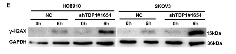
Application: WB Species: human Sample: HO8910 and SKOV3 cells

Application: WB Species: human Sample: IMR-90 cells

Application: IF/ICC Species: human Sample:
Application: WB Species: rat Sample:

Application: IHC Species: rat Sample: brain
Restrictive clause
Affinity Biosciences tests all products strictly. Citations are provided as a resource for additional applications that have not been validated by Affinity Biosciences. Please choose the appropriate format for each application and consult Materials and Methods sections for additional details about the use of any product in these publications.
For Research Use Only.
Not for use in diagnostic or therapeutic procedures. Not for resale. Not for distribution without written consent. Affinity Biosciences will not be held responsible for patent infringement or other violations that may occur with the use of our products. Affinity Biosciences, Affinity Biosciences Logo and all other trademarks are the property of Affinity Biosciences LTD.