製品: Collagen XIV alpha 1 Antibody
カタログ: AF0573
タンパク質の説明: Rabbit polyclonal antibody to Collagen XIV alpha 1
アプリケーション: WB IHC IF/ICC
反応性: Human, Mouse
予測: Bovine, Rabbit, Dog, Xenopus
分子量: 193kDa; 194kD(Calculated).
ユニプロット: Q05707
RRID: AB_2834370

類似製品を見る>>

   サイズ 価格 在庫状況
 100ul $280 在庫あり
 200ul $350 在庫あり

リードタイム: 当日配達

For pricing and ordering contact:
お問い合わせ先

製品説明

ソース:
Rabbit
アプリケーション:
IHC 1:50-1:200, IF/ICC 1:100-1:500, WB 1:500-1:2000
*The optimal dilutions should be determined by the end user.
*Tips:

WB: For western blot detection of denatured protein samples. IHC: For immunohistochemical detection of paraffin sections (IHC-p) or frozen sections (IHC-f) of tissue samples. IF/ICC: For immunofluorescence detection of cell samples. ELISA(peptide): For ELISA detection of antigenic peptide.

反応性:
Human,Mouse
予測:
Bovine(100%), Rabbit(100%), Dog(100%), Xenopus(100%)
クローナリティ:
Polyclonal
特異性:
Collagen XIV alpha 1 Antibody detects endogenous levels of total Collagen XIV alpha 1.
RRID:
AB_2834370
引用形式: Affinity Biosciences Cat# AF0573, RRID:AB_2834370.
コンジュゲート:
Unconjugated.
精製:
The antiserum was purified by peptide affinity chromatography using SulfoLink™ Coupling Resin (Thermo Fisher Scientific).
保存:
Rabbit IgG in phosphate buffered saline , pH 7.4, 150mM NaCl, 0.02% sodium azide and 50% glycerol. Store at -20 °C. Stable for 12 months from date of receipt.
別名:

折りたたみ/展開

Collagen alpha 1(XIV) chain; Collagen type XIV alpha 1 (undulin); Collagen type XIV alpha 1; UND; Undulin (fibronectin tenascin related); Undulin; COL14A1;

免疫原

免疫原:
Uniprot:
遺伝子(ID):
タンパク質の説明:
COL14A1 Plays an adhesive role by integrating collagen bundles. It is probably associated with the surface of interstitial collagen fibrils via COL1. The COL2 domain may then serve as a rigid arm which sticks out from the fibril and protrudes the large N-terminal globular domain into the extracellular space, where it might interact with other matrix molecules or cell surface receptors. Belongs to the fibril-associated collagens with interrupted helices (FACIT) family.
タンパク質配列:
MKIFQRKMRYWLLPPFLAIVYFCTIVQGQVAPPTRLRYNVISHDSIQISWKAPRGKFGGYKLLVTPTSGGKTNQLNLQNTATKAIIQGLMPDQNYTVQIIAYNKDKESKPAQGQFRIKDLEKRKDPKPRVKVVDRGNGSRPSSPEEVKFVCQTPAIADIVILVDGSWSIGRFNFRLVRHFLENLVTAFDVGSEKTRIGLAQYSGDPRIEWHLNAFSTKDEVIEAVRNLPYKGGNTLTGLALNYIFENSFKPEAGSRTGVSKIGILITDGKSQDDIIPPSRNLRESGVELFAIGVKNADVNELQEIASEPDSTHVYNVAEFDLMHTVVESLTRTLCSRVEEQDREIKASAHAITGPPTELITSEVTARSFMVNWTHAPGNVEKYRVVYYPTRGGKPDEVVVDGTVSSTVLKNLMSLTEYQIAVFAIYAHTASEGLRGTETTLALPMASDLLLYDVTENSMRVKWDAVPGASGYLILYAPLTEGLAGDEKEMKIGETHTDIELSGLLPNTEYTVTVYAMFGEEASDPVTGQETTLALSPPRNLRISNVGSNSARLTWDPTSRQINGYRIVYNNADGTEINEVEVDPITTFPLKGLTPLTEYTIAIFSIYDEGQSEPLTGVFTTEEVPAQQYLEIDEVTTDSFRVTWHPLSADEGLHKLMWIPVYGGKTEEVVLKEEQDSHVIEGLEPGTEYEVSLLAVLDDGSESEVVTAVGTTLDSFWTEPATTIVPTTSVTSVFQTGIRNLVVGDETTSSLRVKWDISDSDVQQFRVTYMTAQGDPEEEVIGTVMVPGSQNNLLLKPLLPDTEYKVTVTPIYTDGEGVSVSAPGKTLPSSGPQNLRVSEEWYNRLRITWDPPSSPVKGYRIVYKPVSVPGPTLETFVGADINTILITNLLSGMDYNVKIFASQASGFSDALTGMVKTLFLGVTNLQAKHVEMTSLCAHWQVHRHATAYRVVIESLQDRQKQESTVGGGTTRHCFYGLQPDSEYKISVYTKLQEIEGPSVSIMEKTQSLPTRPPTFPPTIPPAKEVCKAAKADLVFMVDGSWSIGDENFNKIISFLYSTVGALNKIGTDGTQVAMVQFTDDPRTEFKLNAYKTKETLLDAIKHISYKGGNTKTGKAIKYVRDTLFTAESGTRRGIPKVIVVITDGRSQDDVNKISREMQLDGYSIFAIGVADADYSELVSIGSKPSARHVFFVDDFDAFKKIEDELITFVCETASATCPVVHKDGIDLAGFKMMEMFGLVEKDFSSVEGVSMEPGTFNVFPCYQLHKDALVSQPTRYLHPEGLPSDYTISFLFRILPDTPQEPFALWEILNKNSDPLVGVILDNGGKTLTYFNYDQSGDFQTVTFEGPEIRKIFYGSFHKLHIVVSETLVKVVIDCKQVGEKAMNASANITSDGVEVLGKMVRSRGPGGNSAPFQLQMFDIVCSTSWANTDKCCELPGLRDDESCPDLPHSCSCSETNEVALGPAGPPGGPGLRGPKGQQGEPGPKGPDGPRGEIGLPGPQGPPGPQGPSGLSIQGMPGMPGEKGEKGDTGLPGPQGIPGGVGSPGRDGSPGQRGLPGKDGSSGPPGPPGPIGIPGTPGVPGITGSMGPQGALGPPGVPGAKGERGERGDLQSQAMVRSVARQVCEQLIQSHMARYTAILNQIPSHSSSIRTVQGPPGEPGRPGSPGAPGEQGPPGTPGFPGNAGVPGTPGERGLTGIKGEKGNPGVGTQGPRGPPGPAGPSGESRPGSPGPPGSPGPRGPPGHLGVPGPQGPSGQPGYCDPSSCSAYGVRAPHPDQPEFTPVQDELEAMELWGPGV

種類予測

種類予測:

Score>80(red) has high confidence and is suggested to be used for WB detection. *The prediction model is mainly based on the alignment of immunogen sequences, the results are for reference only, not as the basis of quality assurance.

Species
Results
Score
Bovine
100
Dog
100
Xenopus
100
Rabbit
100
Chicken
78
Pig
0
Horse
0
Sheep
0
Zebrafish
0
Model Confidence:
High(score>80) Medium(80>score>50) Low(score<50) No confidence

PTMs - Q05707 基板として

Site PTM Type Enzyme
N94 N-Glycosylation
N372 N-Glycosylation
T439 Phosphorylation
T643 O-Glycosylation
S648 Phosphorylation
Y662 Phosphorylation
S963 Phosphorylation
T970 Phosphorylation
Y988 Phosphorylation
T989 Phosphorylation
T1067 Phosphorylation
T1070 Phosphorylation
T1078 Phosphorylation
Y1118 Phosphorylation
T1122 Phosphorylation
T1125 Phosphorylation
S1128 Phosphorylation
N1384 N-Glycosylation
N1388 N-Glycosylation
S1648 Phosphorylation
S1664 Phosphorylation
S1728 Phosphorylation
S1734 Phosphorylation
Y1758 Phosphorylation
Y1767 Phosphorylation

研究背景

機能:

Plays an adhesive role by integrating collagen bundles. It is probably associated with the surface of interstitial collagen fibrils via COL1. The COL2 domain may then serve as a rigid arm which sticks out from the fibril and protrudes the large N-terminal globular domain into the extracellular space, where it might interact with other matrix molecules or cell surface receptors (By similarity).

PTMs:

Lysines at the third position of the tripeptide repeating unit (G-X-Y) are hydroxylated in all cases and bind carbohydrates.

Prolines at the third position of the tripeptide repeating unit (G-X-Y) are hydroxylated in some or all of the chains.

May contain numerous cysteine residues involved in inter- and intramolecular disulfide bonding.

細胞の位置付け:

Secreted>Extracellular space>Extracellular matrix.

Extracellular region or secreted Cytosol Plasma membrane Cytoskeleton Lysosome Endosome Peroxisome ER Golgi apparatus Nucleus Mitochondrion Manual annotation Automatic computational assertionSubcellular location
サブユニット構造:

Homotrimer.

タンパク質ファミリー:

Belongs to the fibril-associated collagens with interrupted helices (FACIT) family.

研究領域

· Organismal Systems > Digestive system > Protein digestion and absorption.

参考文献

1). Whole-exome mutational landscape and molecular marker study in mucinous and clear cell ovarian cancer cell lines 3AO and ES2. BMC Cancer [IF=3.8]

Application: WB    Species: Human    Sample:

Fig. 6 Differential expression of CCDC170, COL14A1 and THBS2 in ovarian cancer cell lines and normal ovarian epithelial cells. (A) Western blotting was used to detect the expression of CCDC170, COL14A1 and THBS2 in ovarian cancer cell lines and normal ovarian epithelial cells. In order to improve the clarity and conciseness of the presentation, we cropped the blots. The original, untreated blots images can be found in Additional file 6. (B-D) Statistical analysis of Western blotting results of CCDC170, COL14A1 and THBS2 proteins. (E-G) The expression of CCDC170, COL14A1 and THBS2 in ovarian cancer cell lines and normal ovarian epithelial cells was detected by immunofluorescence assay. Original magnification: 40×. (H-J) Statistical analysis of mean fluorescence density of CCDC170, COL14A1 and THBS2 proteins. The gray values of protein bands and the fluorescence intensity of immunofluorescence were analyzed using Image J software (National Institutes of Health, Bethesda, MD, USA).

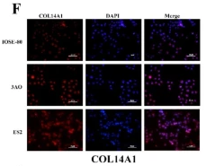
Application: IF/ICC    Species: Human    Sample:

Fig. 6 Differential expression of CCDC170, COL14A1 and THBS2 in ovarian cancer cell lines and normal ovarian epithelial cells. (A) Western blotting was used to detect the expression of CCDC170, COL14A1 and THBS2 in ovarian cancer cell lines and normal ovarian epithelial cells. In order to improve the clarity and conciseness of the presentation, we cropped the blots. The original, untreated blots images can be found in Additional file 6. (B-D) Statistical analysis of Western blotting results of CCDC170, COL14A1 and THBS2 proteins. (E-G) The expression of CCDC170, COL14A1 and THBS2 in ovarian cancer cell lines and normal ovarian epithelial cells was detected by immunofluorescence assay. Original magnification: 40×. (H-J) Statistical analysis of mean fluorescence density of CCDC170, COL14A1 and THBS2 proteins. The gray values of protein bands and the fluorescence intensity of immunofluorescence were analyzed using Image J software (National Institutes of Health, Bethesda, MD, USA).

2). Identification of Underlying Hub Genes Associated with Hypertrophic Cardiomyopathy by Integrated Bioinformatics Analysis. Pharmacogenomics and Personalized Medicine (PubMed: 34285551)

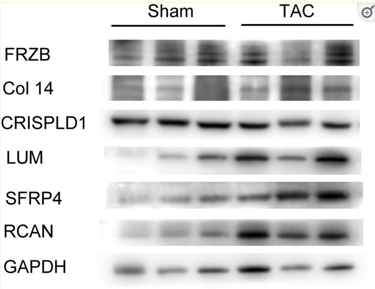
Application: WB    Species: Mouse    Sample: myocardium

Figure 8 Western blotting tests representing protein levels of hub genes (FRZB, COL14A1, CRISPLD1, LUM, sFRP4) and remodeling gene RCAN in myocardium of sham and TAC mice. Abbreviations: FRZB, frizzled related protein; COL14A1, collagen type XIV alpha 1 chain; CRISPLD1, cysteine rich secretory protein LCCL domain containing 1; LUM, lumican; sFRP4, secreted frizzled related protein 4; RCAN, calcipressin regulatory protein.

Restrictive clause


Affinity Biosciences tests all products strictly. Citations are provided as a resource for additional applications that have not been validated by Affinity Biosciences. Please choose the appropriate format for each application and consult Materials and Methods sections for additional details about the use of any product in these publications.

For Research Use Only.
Not for use in diagnostic or therapeutic procedures. Not for resale. Not for distribution without written consent. Affinity Biosciences will not be held responsible for patent infringement or other violations that may occur with the use of our products. Affinity Biosciences, Affinity Biosciences Logo and all other trademarks are the property of Affinity Biosciences LTD.