, using DNER Antibody at 1/1000 dilution.
5ug/NC membrane strip.
Exposure for 1min with Affinity™ ECL Kit(#KF8003).
Bands result from membrane strip incubation.")


製品説明
*The optimal dilutions should be determined by the end user. For optimal experimental results, antibody reuse is not recommended.
*Tips:
WB: For western blot detection of denatured protein samples. IHC: For immunohistochemical detection of paraffin sections (IHC-p) or frozen sections (IHC-f) of tissue samples. IF/ICC: For immunofluorescence detection of cell samples. ELISA(peptide): For ELISA detection of antigenic peptide.
引用形式: Affinity Biosciences Cat# DF10181, RRID:AB_2840760.
折りたたみ/展開
bet; Brain EGF repeat-containing transmembrane protein; Bret; Delta and Notch-like epidermal growth factor-related receptor; Delta notch like EGF repeat containing transmembrane; Delta/notch like EGF repeat containing; Delta/notch-like EGF-related receptor; Dner; DNER_HUMAN; PRO299; Transmembrane protein Bet; UNQ26;
免疫原
A synthesized peptide derived from human DNER, corresponding to a region within the internal amino acids.
- Q8NFT8 DNER_HUMAN:
- Protein BLAST With
- NCBI/
- ExPASy/
- Uniprot
MQPRRAQAPGAQLLPALALLLLLLGAGPRGSSLANPVPAAPLSAPGPCAAQPCRNGGVCTSRPEPDPQHPAPAGEPGYSCTCPAGISGANCQLVADPCASNPCHHGNCSSSSSSSSDGYLCICNEGYEGPNCEQALPSLPATGWTESMAPRQLQPVPATQEPDKILPRSQATVTLPTWQPKTGQKVVEMKWDQVEVIPDIACGNASSNSSAGGRLVSFEVPQNTSVKIRQDATASLILLWKVTATGFQQCSLIDGRSVTPLQASGGLVLLEEMLALGNNHFIGFVNDSVTKSIVALRLTLVVKVSTCVPGESHANDLECSGKGKCTTKPSEATFSCTCEEQYVGTFCEEYDACQRKPCQNNASCIDANEKQDGSNFTCVCLPGYTGELCQSKIDYCILDPCRNGATCISSLSGFTCQCPEGYFGSACEEKVDPCASSPCQNNGTCYVDGVHFTCNCSPGFTGPTCAQLIDFCALSPCAHGTCRSVGTSYKCLCDPGYHGLYCEEEYNECLSAPCLNAATCRDLVNGYECVCLAEYKGTHCELYKDPCANVSCLNGATCDSDGLNGTCICAPGFTGEECDIDINECDSNPCHHGGSCLDQPNGYNCHCPHGWVGANCEIHLQWKSGHMAESLTNMPRHSLYIIIGALCVAFILMLIILIVGICRISRIEYQGSSRPAYEEFYNCRSIDSEFSNAIASIRHARFGKKSRPAMYDVSPIAYEDYSPDDKPLVTLIKTKDL
種類予測
Score>80(red) has high confidence and is suggested to be used for WB detection. *The prediction model is mainly based on the alignment of immunogen sequences, the results are for reference only, not as the basis of quality assurance.
High(score>80) Medium(80>score>50) Low(score<50) No confidence
研究背景
Activator of the NOTCH1 pathway. May mediate neuron-glia interaction during astrocytogenesis (By similarity).
Cell membrane>Single-pass type I membrane protein.
Note: Present on the membrane of dendrites and cell bodies but excluded from axonal membrane. Also found in early endosomes in the somatodendritic region (By similarity).
Expressed in brain, spinal cord and adrenal gland.
参考文献
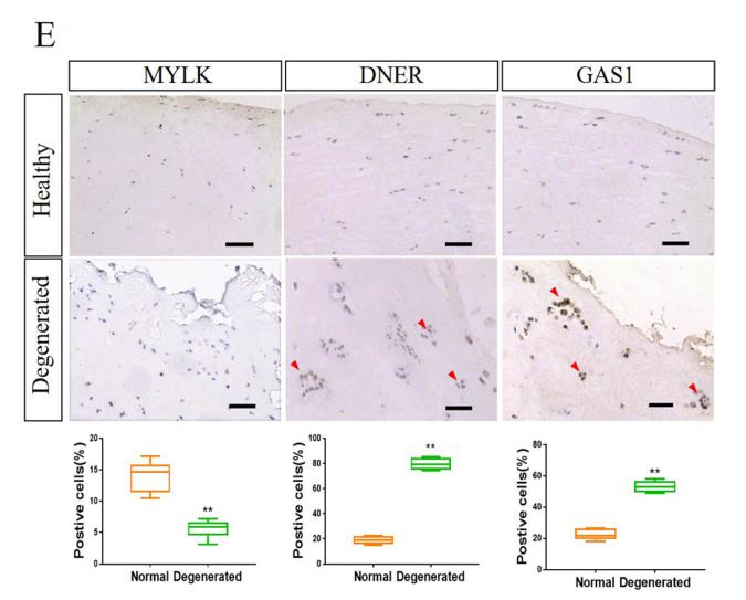
Application: IHC Species: Human Sample: human meniscus cells
Restrictive clause
Affinity Biosciences tests all products strictly. Citations are provided as a resource for additional applications that have not been validated by Affinity Biosciences. Please choose the appropriate format for each application and consult Materials and Methods sections for additional details about the use of any product in these publications.
For Research Use Only.
Not for use in diagnostic or therapeutic procedures. Not for resale. Not for distribution without written consent. Affinity Biosciences will not be held responsible for patent infringement or other violations that may occur with the use of our products. Affinity Biosciences, Affinity Biosciences Logo and all other trademarks are the property of Affinity Biosciences LTD.